Figure 7. Retinal artery tumor necrosis factor (TNF)-R1 immunofluorescence. Images show representative examples of TNF-R1 immunofluorescence
staining in an artery from (A) a sham-operated eye and (B) the fellow eye subjected to ischemia and 12 h of reperfusion. C-E: Enlargements show double staining for (C-E) TNF-R1 and (E) the smooth muscle cell marker, smooth muscle actin (SMA). It can be shown that TNF-R1 and SMA co-localize in the smooth
muscle cells in the blood vessels. D-E: Furthermore, staining with DAPI (which labels the cell nuclei) showed that TNF-R1 was located in the cell membrane of the
smooth muscle cells. The different images are from separate sections.
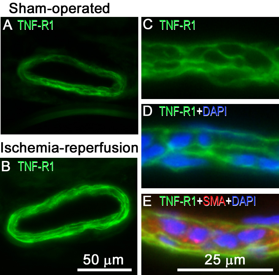
 Figure 7 of
Gesslein, Mol Vis 2010; 16:2317-2327.
Figure 7 of
Gesslein, Mol Vis 2010; 16:2317-2327.