Figure 5. Tumor necrosis factor α (TNF-α) immunofluorescence in the neuroretina. TNF-α immunofluorescence staining was detected in the
outer plexiform layer and in the Müller cells of the neuroretina in (A) a sham-operated eye and (B) the fellow eye subjected to ischemia and 12 h of reperfusion. C-D: Enlargement showing TNF-α staining in the (C) Müller cell processes (arrow heads) and (D) cellbodies of the outer plexiform layer (arrow). E: Enlargement showing double staining with TNF-α and calbindin (a horizontal cell marker) and (F) TNF-α with its receptor TNF receptor 1 (TNF-R1). DAPI was used to label the cell nuclei. The different images are from separate
sections of the retina. Abbreviations used in the figure are outer nuclear layer (ONL), inner nuclear layer (INL), ganglion
cell layer (GCL), and outer plexiform layer (OPL).
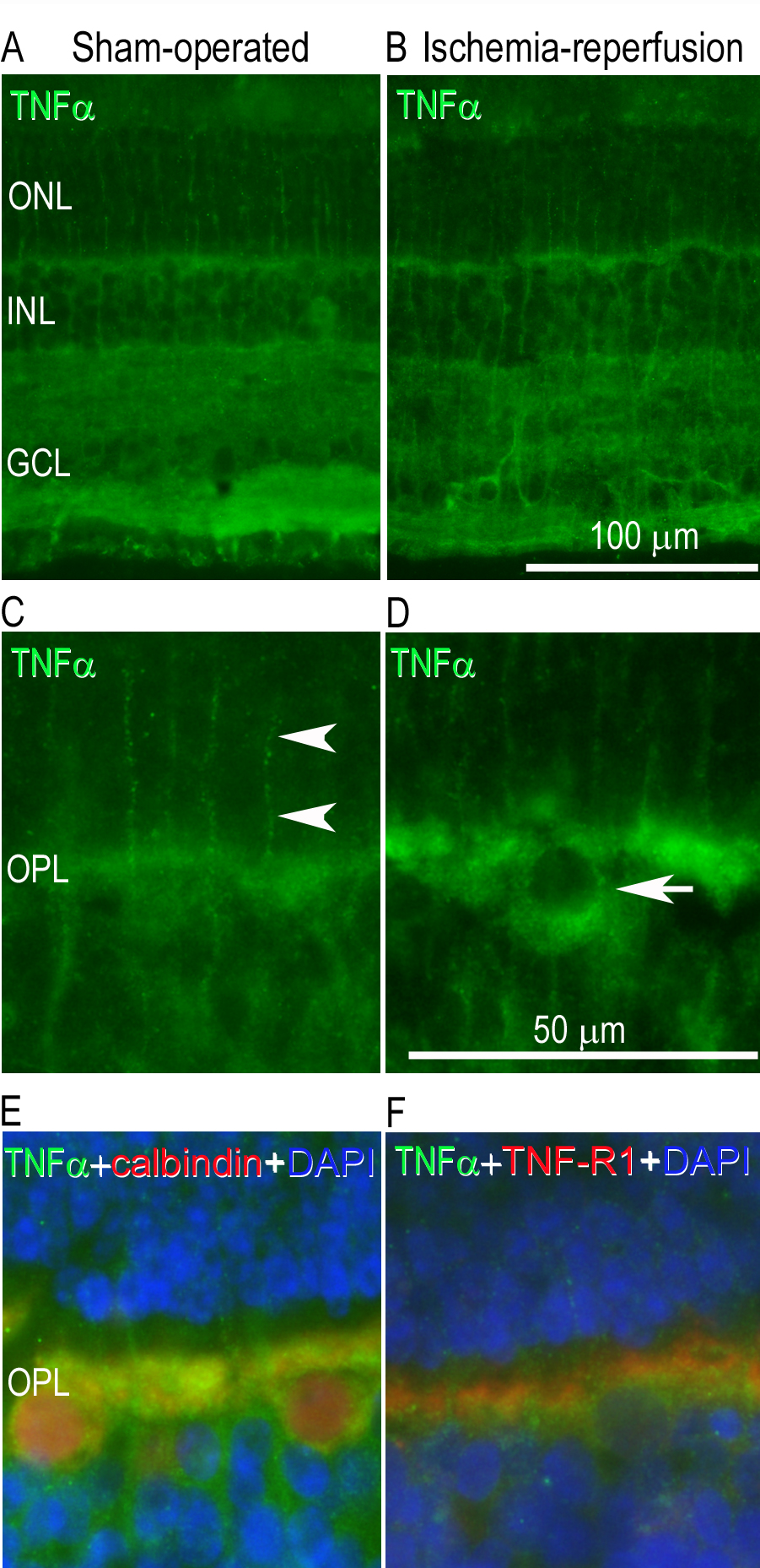
 Figure 5 of
Gesslein, Mol Vis 2010; 16:2317-2327.
Figure 5 of
Gesslein, Mol Vis 2010; 16:2317-2327.