Figure 1. Fundus images from an eye
subjected to successively increasing intraocular pressure. At normal
pressure (10–20 mmHg) the pigs had a normal fundus with filled
blood vessels. The filling of the blood vessels decreased and the
fundus became paler as the intraocular pressure (IOP) increased. At an
IOP of 80 mmHg, blood flow was completely inhibited. IOP is given
with ±5 mmHg variability.
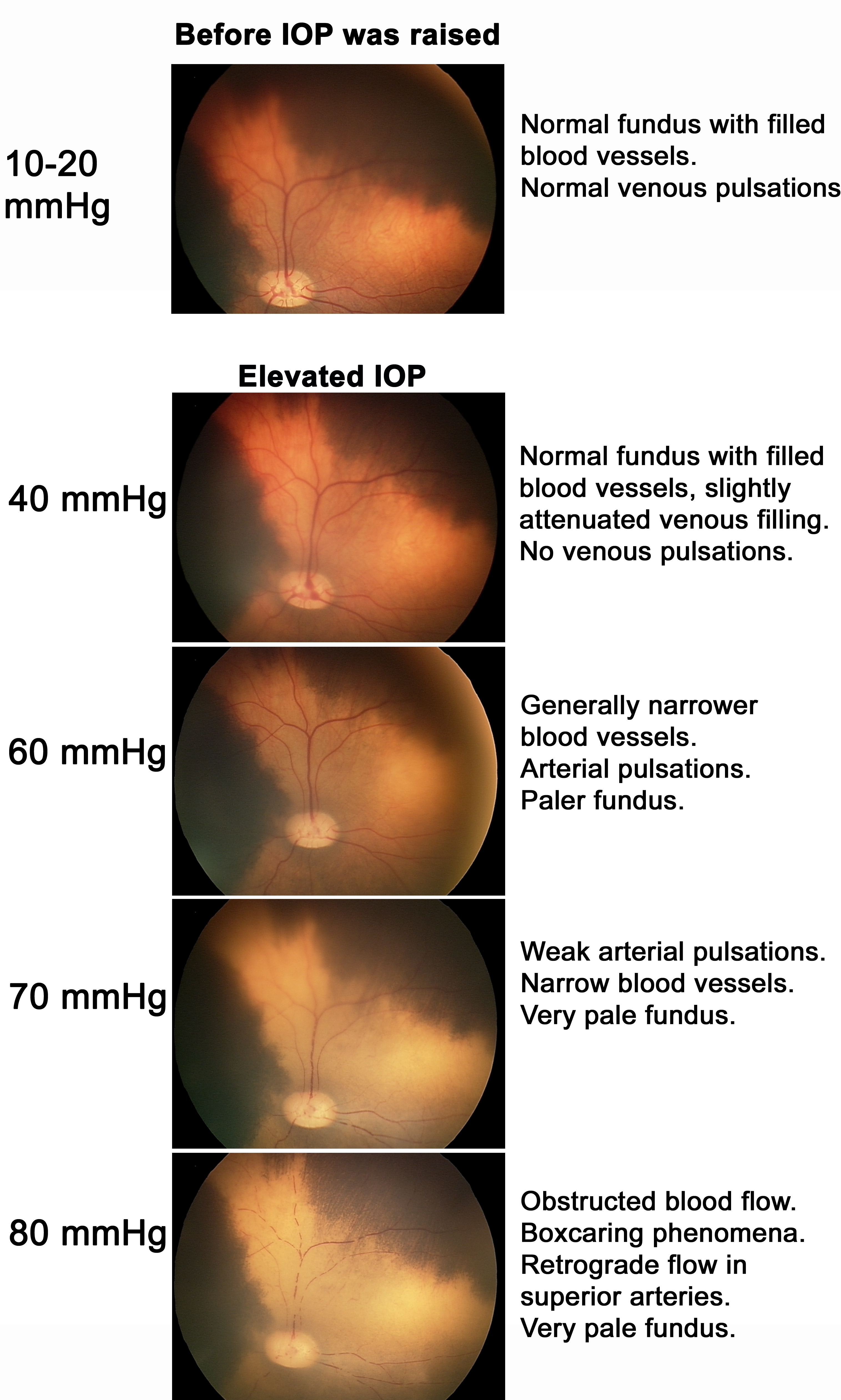
 Figure 1 of Gesslein, Mol Vis 2010; 16:2317-2327.