Figure 7. HuR is expressed in progenitor
lens epithelial cells, and neuronal HuB/C/D in post-mitotic elongating
fiber cells. A: Amplification of HuB, HuC, and HuD transcripts
from lens and brain; right: Region from sequenced product showing in
frame exon junctions in amplified transcripts. B: Amplification
of the HuD target transcripts: GAP43 and CamKIIα from wt mouse and rat
lens and brain. C: Immunoblot detection of HuR in lens and
brain. D: Immunoblot of HuB/C/D in lens and brain (Human
anti-HuB/C/D). E: GAP43 protein detected in wt lens and brain. F:
Increased
expression
of
GAP43 and CamKIIα detected on immunoblots of wt
versus transgenic mouse lenses expressing myc-tagged HuD in the lens;
unchanged GAPDH levels are shown for comparison. G:
Immunofluorescence detection in rat lens: H: mAb anti-HuR
(200×), I: DAPI stained nuclei as in panel H (100×), J:
Human
anti-HuB/C/D,
K: DAPI stained nuclei as in panel J
(200×), L: Human anti-HuB/C/D (100×), M:
anti-COOH-terminal REST, N: overlay of panels L and M,
O: Syn1 expression in post-mitotic fiber cells (BD Biosciences,
200×), P: GAP43 in wt lens, Q: DAPI stained nuclei as
in panel P, R: CamKIIα in wt lens, S: mAb
anti-HuD detection in wt mouse lens (L-P, Santa Cruz).
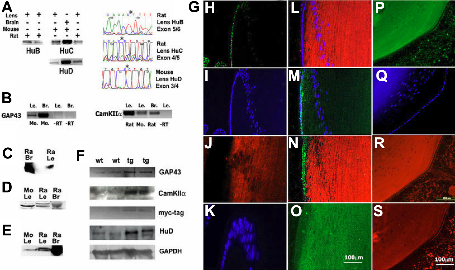
 Figure 7 of Bitel, Mol Vis 2010; 16:2301-2316.