Figure 6. PTB (PTBP1) is expressed in
progenitor epithelial cell and neuronal nPTB (PTBP2) in post-mitotic
lens fiber cells. A: Immunoblot detection of PTB and nPTB in
mouse and rat lens and brain tissue. PTB-NT: anti-NH2-terminus
PTB,
PTB-CT: anti-COOH-terminus PTB, and anti-nPTB. B: Left:
Amplified PTB and nPTB transcripts from lens and brain. Right: Region
from amplified DNA sequence product identifying in frame exon junctions
in lens. C: Immunofluorescence detection of PTB and nPTB in the
lens. D: anti-nPTB (100×), E: anti-PTB-NT (100×), F:
anti-PTB-CT
(100×), G: DAPI nuclear stain/no 1o control (200×),
H: anti-nPTB (200×), I: overlay panel G and H.
J: For comparison PTB is detected in cell nuclei in rat skin, K:
anti-nPTB
detects little or no protein in rat skin, L:
Detection of PTB in cell nuclei in epithelial cells on the anterior
lens surface.
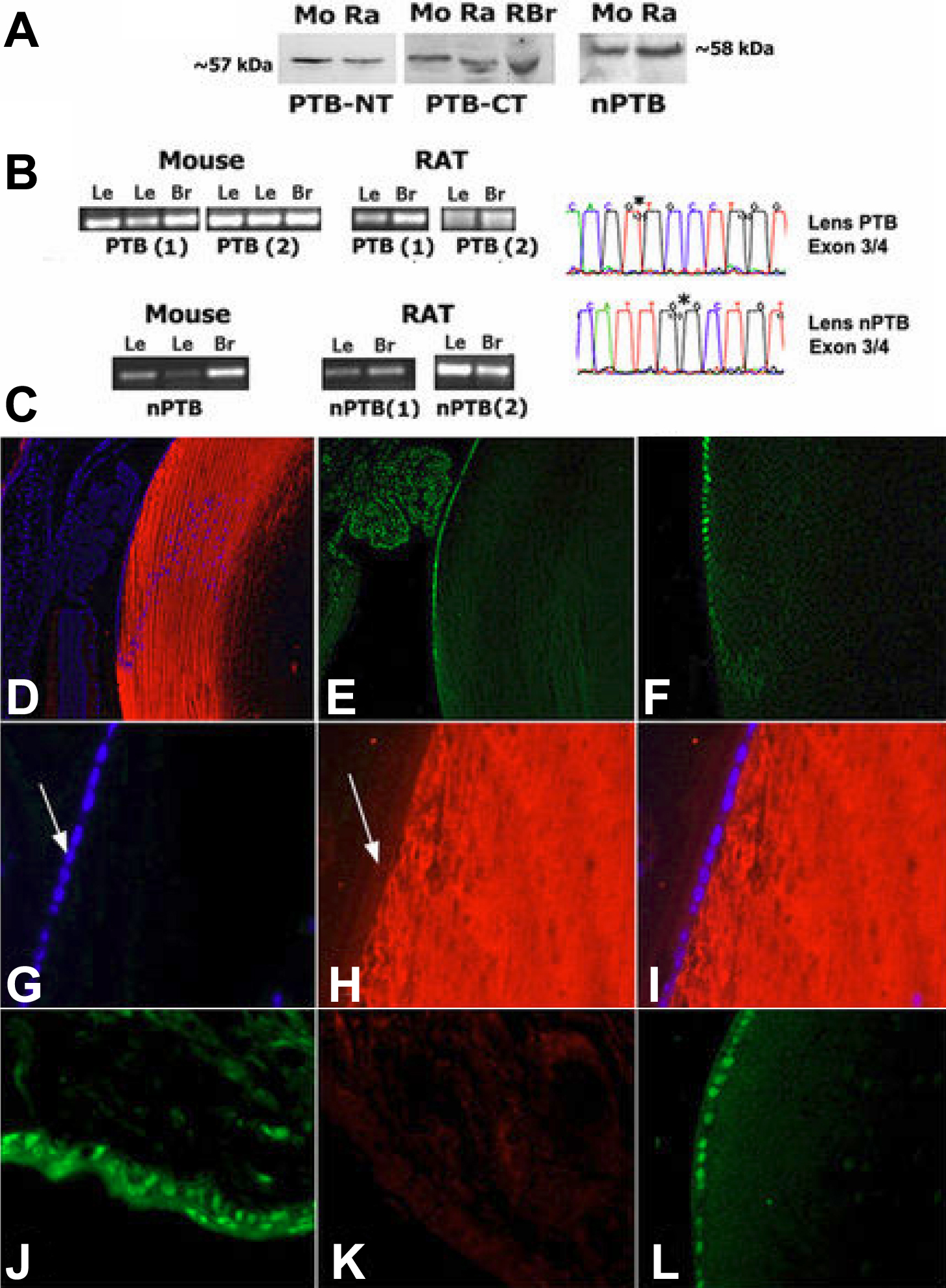
 Figure 6 of Bitel, Mol Vis 2010; 16:2301-2316.