Figure 3. REST regulated neuronal genes
are activated in post-mitotic fiber cells. A: Hematoxylin and eosin
stained mouse lens. B: Mouse mAb βIII-tubulin. C:
Rabbit mAb anti-βIII-tubulin. D: Chicken anti-βIII-tubulin. E:
Anti-Synaptophysin
1. F: Mouse mAb Synaptotagmin 1. G:
Rabbit anti-Synapsin 1. H: DAPI as in G. I: No
primary antibody control (lo), DAPI. J: Synapsin 1 in retinal
neuronal layers (autofluorescence is seen in photoreceptor layer). K:
mAb
βIII-tubulin. L: Synaptotagmin 1 in peripheral fiber cells.
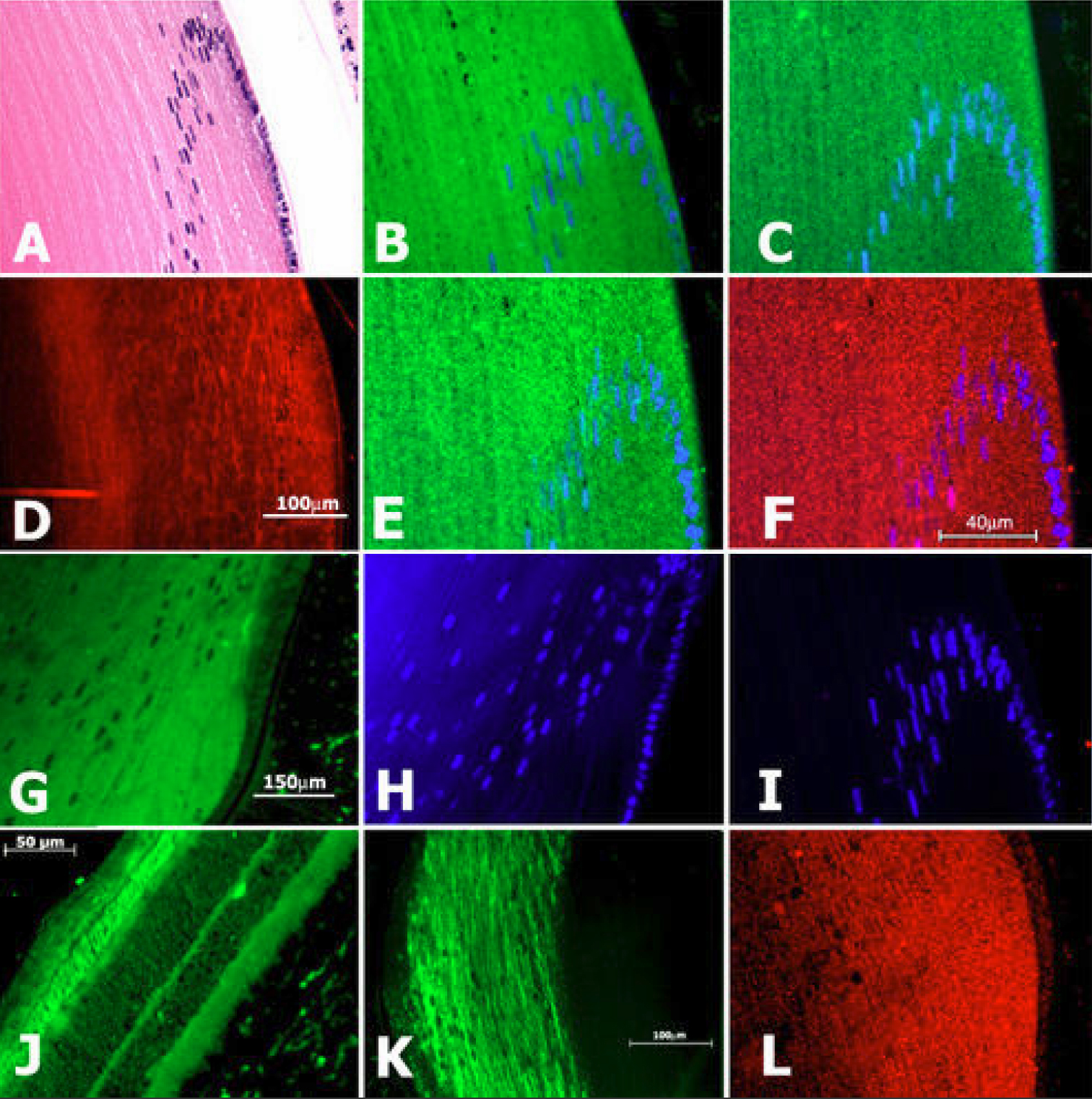
 Figure 3 of Bitel, Mol Vis 2010; 16:2301-2316.