Figure 1. Glass-bottomed chamber for
positioning and viewing mouse lenses. Oblique view (A) and a
plan view (B). The base of the chamber contains a layer of
solidified agar from which a wedge-shaped piece has been removed. To
image the anterior pole (P), the lens, resting on its epithelium, is
positioned in the base of the wedge. To visualize the equatorial region
(E), the lens is turned on its side and positioned such that its
anterior and posterior faces are supported by the gently tapering walls
of the wedge and the equatorial cells are exposed.
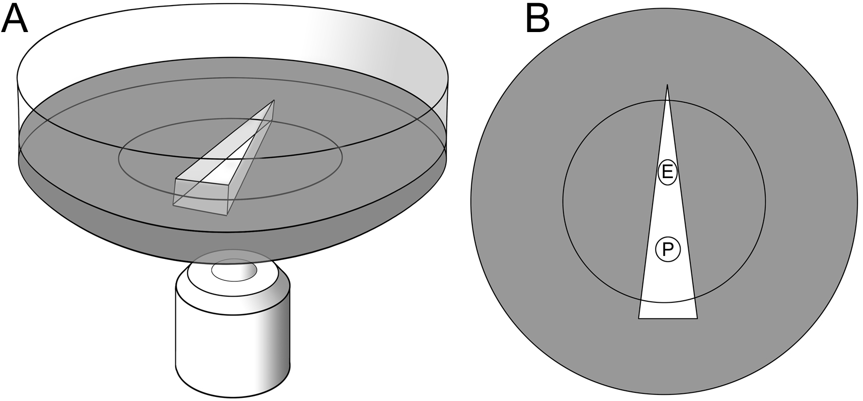
 Figure 1 of Bassnett, Mol Vis 2010; 16:2294-2300.