Figure 1. Expression of ENaC subunits in
rabbit conjunctiva in immunohistochemical stainnings. ENaC-α (A
and I), ENaC-β (B and J), and ENaC-γ (C
and K) were positive at apical layer of conjunctiva. As a
positive control, ENaC-α (E and M), ENaC-β (F
and N), and ENaC-γ (G and O) were positive
at inner medullas of kidney. Isotype control specimen showed no
positive staining in conjunctiva (D and L) and kidney (H
and P). Scale bars in A-H represent 20 μm, and
scale bars in I-P represent 40 μm.
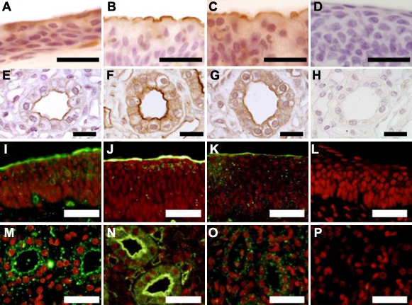
 Figure 1 of Hara, Mol Vis 2010; 16:2279-2285.