Figure 5. Localization of plasma membrane calcium-ATPase (PMCA) in rabbit corneal epithelium (CE). A: Immunostaining with pan-PMCA antibody revealed strong PMCA labeling in all layers of the CE. The staining was associated
primarily with the plasma membranes with diffuse cytoplasmic staining in some cells. The corneal stroma (S) was unstained.
B: The pattern and intensity of immunoreactivity seen with PMCA4 isoform-specific antibody was similar to that seen with the
panPMCA antibody, except that PMCA4 labeling is absent (arrows) from most basal cell plasma membranes adjacent to the stroma.
C: Control section incubated with nonimmune mouse IgG revealed an absence of staining.
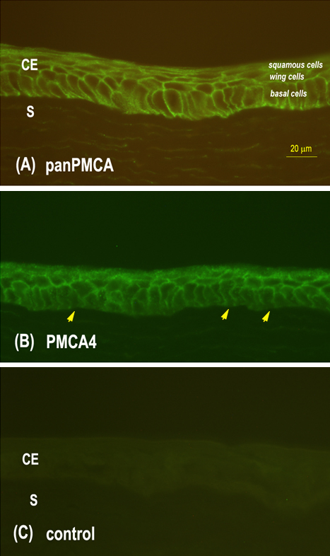
 Figure 5 of
Talarico, Mol Vis 2010; 16:2259-2272.
Figure 5 of
Talarico, Mol Vis 2010; 16:2259-2272.