Figure 4. Immunoblot analysis of plasma membrane calcium-ATPase (PMCA) in rabbit corneal epithelium. Blot lanes were loaded with equal
aliquots of whole cell lysate. The panPMCA antibody was used at 0.65 mg/ml, and the PMCA4 antibody was used at 0.4 mg/ml (data
presented here is from a single animal and is representative of immunoblot studies of the CE from three different normal animals).
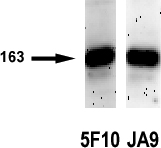
 Figure 4 of
Talarico, Mol Vis 2010; 16:2259-2272.
Figure 4 of
Talarico, Mol Vis 2010; 16:2259-2272.