Figure 2. Redistribution of plasma membrane calcium-ATPase isoform 4 (PMCA4) in basal epithelial cells at the wound margin during corneal
reepithelialization. The pattern of PMCA4 IR is shown in the wound margin at 6 h (A), 24 h (B), 36 h (C), and 48 h (D), and in the control cornea at 6 h (E). The presence (asterisks) or absence (arrows) of basal plasma membrane staining in representative cells is shown (corneal
epithelium [CE]; stroma [S]). Bars in A represent 20 µm and apply to all panels.
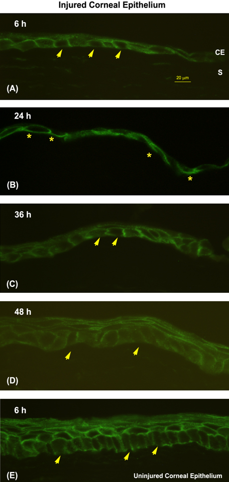
 Figure 2 of
Talarico, Mol Vis 2010; 16:2259-2272.
Figure 2 of
Talarico, Mol Vis 2010; 16:2259-2272.