Figure 2. Immunostaining for S100A8, A9, and cellular markers in murine corneas. Neutrophils and macrophages were stained green via
primary rabbit anti-Gr-1 (A) or anti-F4/80 (B) and FITC-conjugated secondary antibodies, while S100A8 or A9 were stained red via PE conjugated primary antibodies. Please
note that the autofluorescence for corneal epithelium should not be misinterpreted as positive staining.
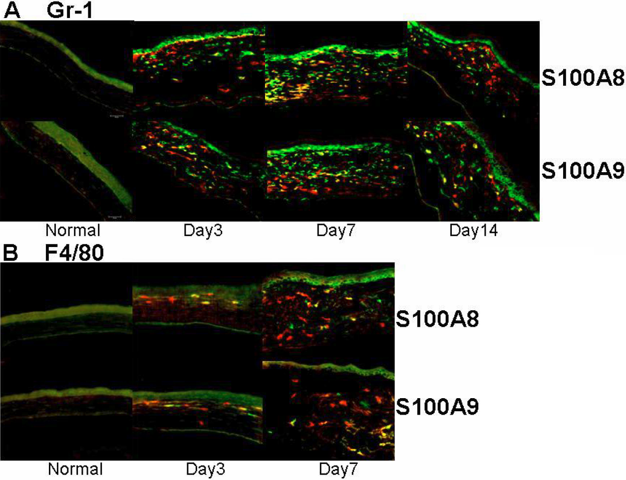
 Figure 2 of
Li, Mol Vis 2010; 16:2225-2235.
Figure 2 of
Li, Mol Vis 2010; 16:2225-2235.