Figure 3. PALM laser dissection. A, D, G: OCT-embedded mouse eye tissue was cut at 10 μm, fixed and stained with hematoxylin and Eosin. B, E, H: Palpebral, bulbar and forniceal conjunctival epithelium region were identified and circled. C, F, I: The selected region was cut from the surrounding cells, and the same region was captured leaving a clear margin of surrounding
cells. All pictures were taken at 400×. bcj, bulbar conjunctiva; pcj, palpebral conjunctiva.
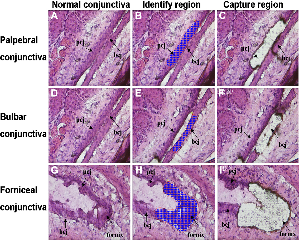
 Figure 3 of
Zhu, Mol Vis 2010; 16:2215-2224.
Figure 3 of
Zhu, Mol Vis 2010; 16:2215-2224.