Figure 1. Distribution of microfilament regulators in the conjunctiva. bcj, bulbar conjunctiva; pcj, palpebral conjunctiva. A: Integrin β1 (green) and CK4 (Red); B: Integrin α6 (green) and CK4 (red); C: Talin1 (green); D: Vinculin (green); E: Profilin1 (green); F: Double staining of profilin1 (red) and phalloidin (green); G: Paxillin (green); H: Double staining of paxillin (red) and phalloidin (green); I: FAK (green); J: Double staining of FAK (red) and phalloidin (green); K: Cofilin1 (green); L: Double staining of cofilin1 (red) and phalloidin (green); M: Gelsolin (green color); N: Double staining of gelsolin (red) and phalloidin (green); P, Q: Integrin β1 (green); R, S: Paxillin (green); T, U: Profilin1 (green). Blue color is DAPI as a counterstain, staining nuclear.
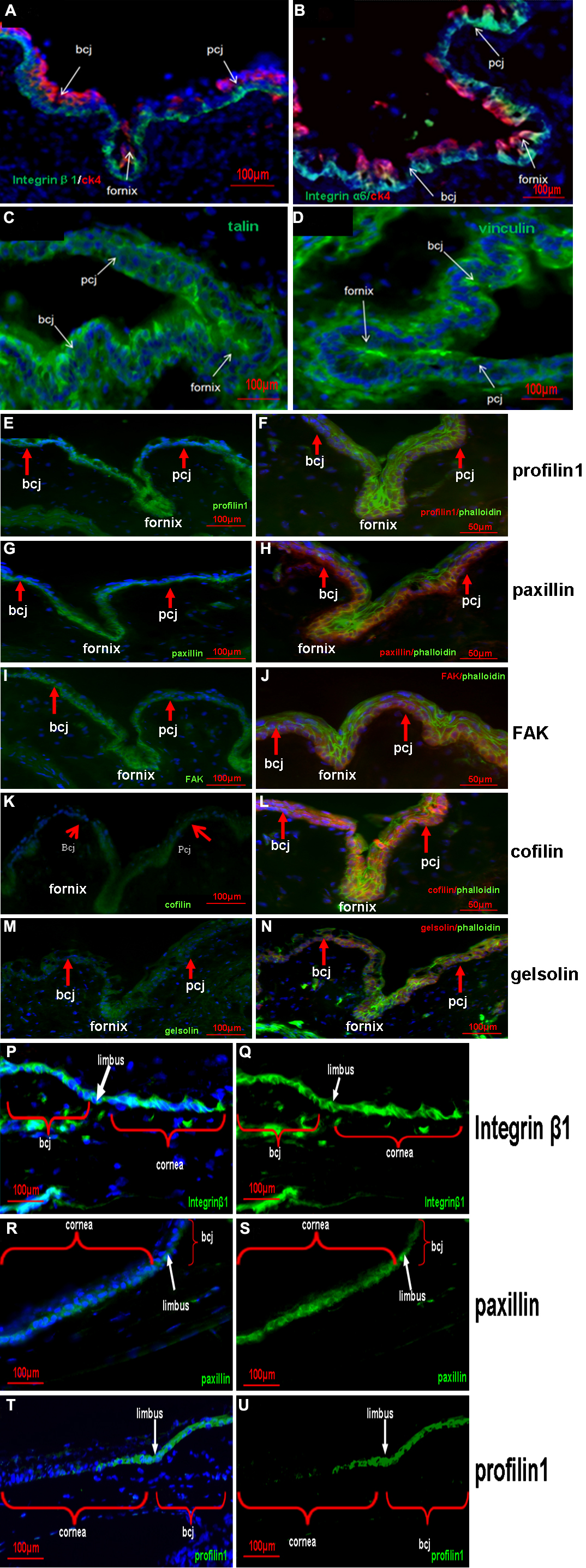
 Figure 1 of
Zhu, Mol Vis 2010; 16:2215-2224.
Figure 1 of
Zhu, Mol Vis 2010; 16:2215-2224.