Figure 3. AR-12 treatment in the ex vivo
model of PCO following 14 days. A: Lens capsules treated with
DMEM media and vehicle as viewed by phase contrast microscopy (scale
bar is equal to 20 µm); the cells are healthy and cuboidal to hexagonal
in shape with very little anisocytosis. B: Lens capsules
treated with DMEM and vehicle (H&E, scale bar is equal to 30 µm);
LEC are of normal morphology and present across the anterior and
posterior lens capsules. C: Lens capsules treated with 2.5µM
AR-12 and viewed by phase contrast microscopy (scale bar is equal to 20
µm); the cells are mostly cuboidal to hexagonal in shape but some
vacuolation is present. D: Lens capsules treated with 2.5 µM
AR-12 (H&E, scale bar is equal to 30 µm); LEC display anisocytosis
and are fewer in number compared to control. E: Phase contrast
photograph (scale bar is equal to 20 µm) or F: H&E
sectioning (scale bar is equal to 30 µm) of lens capsules treated with
5 µM AR-12; there is marked vacuolation, cells lack normal morphology
and there are areas of capsule devoid of epithelial cells. Lens
capsules treated with 7.5 µM AR-12 as viewed by either G: phase
contrast microscopy (scale bar is equal to 20 µm) or H: H&E
sectioning (scale bar is equal to 50 µm) had no normal LEC; few cells
and cellular debris remains within the lens capsule and are adherent to
the anterior or posterior lens capsules. I: Lens capsules
treated with 10 µM AR-12 and viewed by phase contrast microscopy (scale
bar is equal to 20 µm); the lens capsule is devoid of LEC. J:
Lens capsules treated with 10 µM AR-12 (H&E, scale bar is equal to
30 µm); anterior and posterior lens capsules are without LEC.
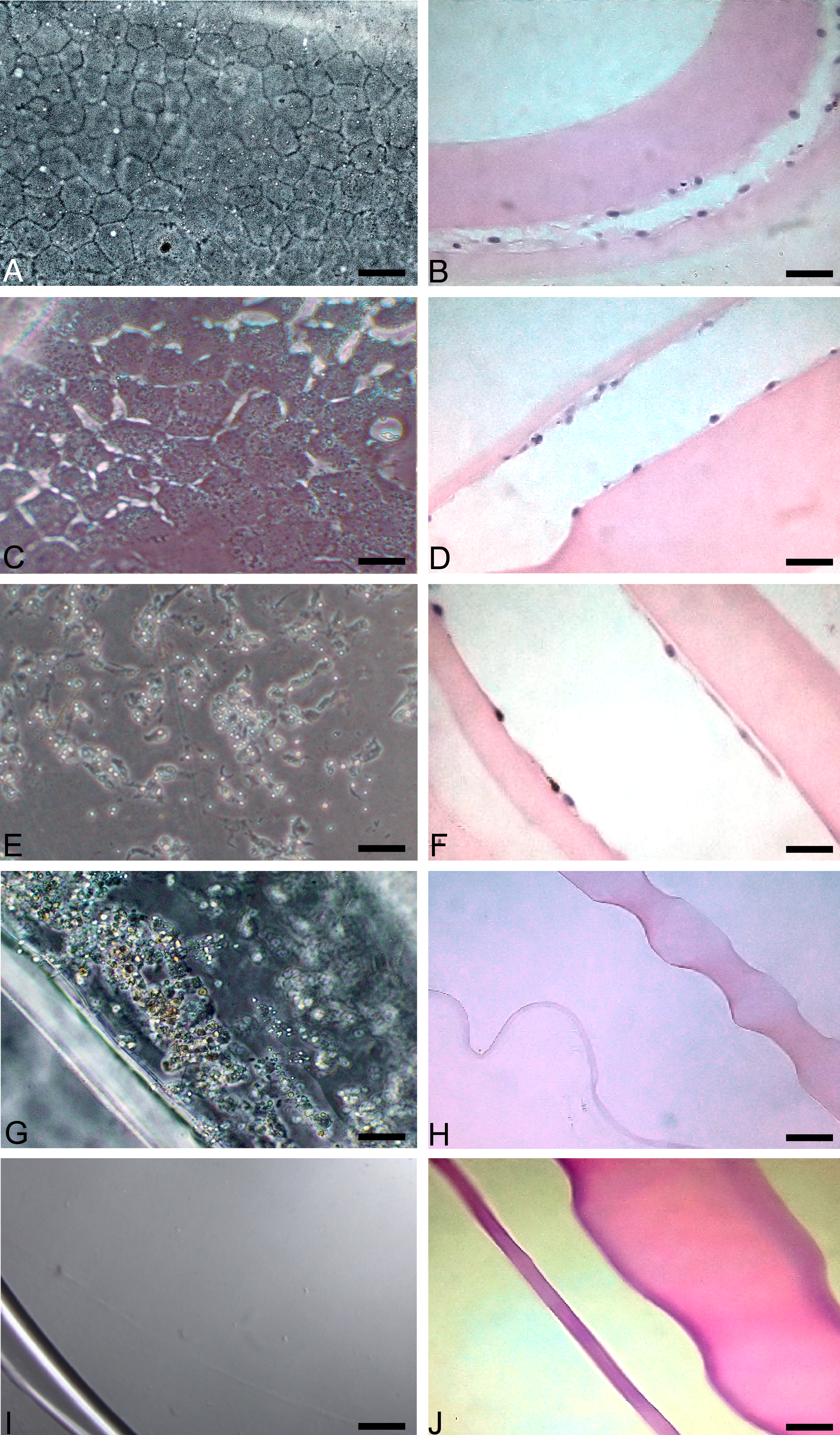
 Figure 3 of Chandler, Mol Vis 2010; 16:2202-2214.