Figure 1. Expression of pAkt in normal canine LEC and in LEC undergoing EMT. A: In normal LEC there is little to no immunoreactivity, while LEC in B: clinical cataracts and C: clinical samples of PCO, show marked cytoplasmic staining for pAkt. The scale bar is equal to 30 µm. D: western blot analysis demonstrates increased expression of pAkt protein in both cataractous LEC and LEC in the ex vivo model
of PCO formation, compared to normal LEC. The ‘+’ indicates a canine mast cell tumor which was used as a positive control
for pAkt expression.
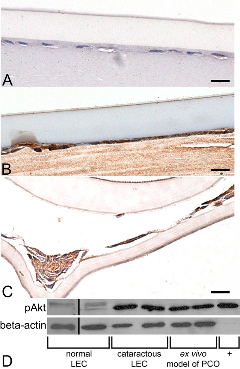
 Figure 1 of
Chandler, Mol Vis 2010; 16:2202-2214.
Figure 1 of
Chandler, Mol Vis 2010; 16:2202-2214.