Figure 7. Immunofluorescence staining of Na+/K+-ATPase α1. A, C: The corneal epithelium has a bottom-up decreasing gradient of expression of the Na+/K+-ATPase pumps (in red) from the basal layer toward the wing cells in both native corneas (A) and reconstructed corneas (C). B, D: Immunofluorescence staining of Na+/K+-ATPase α1 of the corneal endothelium in both native (B) and reconstructed corneas (D). Nuclei were counterstained with Hoechst (in blue). Bar, 10 µm.
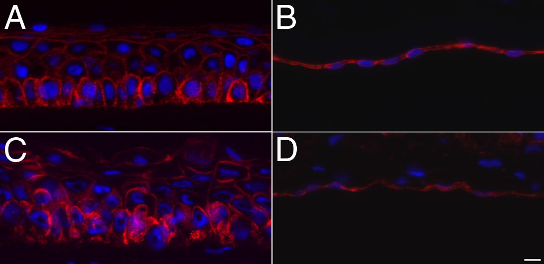
 Figure 7 of
Proulx, Mol Vis 2010; 16:2192-2201.
Figure 7 of
Proulx, Mol Vis 2010; 16:2192-2201.