Figure 6. Epithelial basement membrane components. A, B: Native human corneas. C, D: Tissue-engineered corneas. A, C: Immunofluorescence staining of collagen VII (in red). B, D: Immunofluorescence staining of laminin V (in red). Nuclei were counterstained with Hoechst (in blue). Bar, 10 µm.
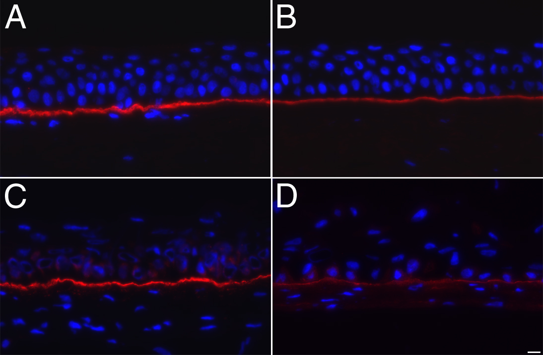
 Figure 6 of
Proulx, Mol Vis 2010; 16:2192-2201.
Figure 6 of
Proulx, Mol Vis 2010; 16:2192-2201.