Figure 4 of
Proulx, Mol Vis 2010; 16:2192-2201.
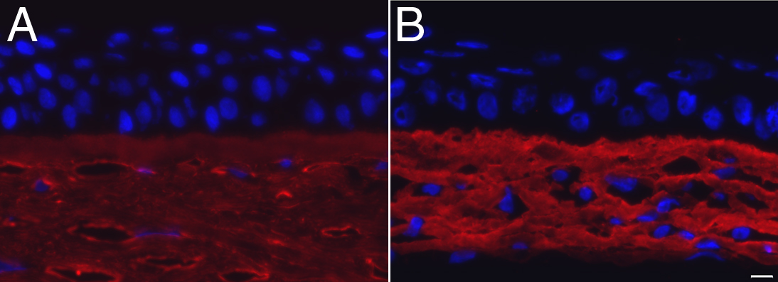
Figure 4.
Collagen type I immunofluorescence staining. Strong collagen type I staining (in red) was found in both the native (
A
) and reconstructed stromas (
B
). Nuclei were counterstained with Hoechst (in blue). Bar, 10 µm.
 Figure 4 of
Proulx, Mol Vis 2010; 16:2192-2201.
Figure 4 of
Proulx, Mol Vis 2010; 16:2192-2201.