Figure 3. Tissue-engineered human cornea. A: Histology (Masson’s Trichrome staining) of the tissue-engineered cornea, showing a well differentiated epithelium on top,
and a monolayer of endothelial cells underneath, both adhered to the self-assembled stromal matrix. B: Transmission electron microscopy of the epithelial basal membrane showing many hemidesmosomes (arrows). C: Transmission electron microscopy of the corneal endothelium, showing a monolayer of flattened cells. The bar in A equals 50 µm, in B equals 1 µm, and in C equals 0.5 µm.
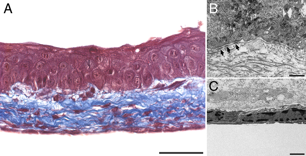
 Figure 3 of
Proulx, Mol Vis 2010; 16:2192-2201.
Figure 3 of
Proulx, Mol Vis 2010; 16:2192-2201.