Figure 1. Protein profiles of the posterior sclera on 2-DE. The 2-DE distributing profile of the differentially expressed protein spots
from the posterior sclera distinct between the normal control eyes and the MD eyes or the recovery eyes (A). Synthetic gel produced from triplicate gels of normal control eyes (B), the differentially expressed protein spots were framing labeled.
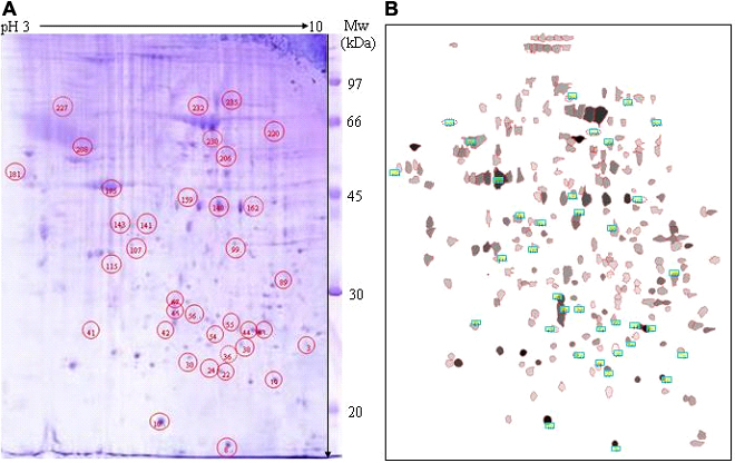
 Figure 1 of
Zhou, Mol Vis 2010; 16:2163-2174.
Figure 1 of
Zhou, Mol Vis 2010; 16:2163-2174.