Figure 6. Level of IL-4 and HSV-1 gB
transcripts in RS cells infected with different recombinant viruses.
Subconfluent monolayers of RS cells were infected with 10 PFU/cell of
HSV-IL-4, vEye2, vEye3, vTG2, or vTG3. Total RNA was isolated 24 and
48hr post infection and TaqMan RT–PCR was performed using IL-4-
and gB-specific primers as described in the Methods. In each
experiment, an estimated relative copy number of IL-4 and gB
were calculated using standard curves generated from pVR1055-IL-4 and
pVR1055-gB, respectively. Briefly, DNA template was serially diluted
10-fold such that 5 μl contained from 103 to 1011
copies of IL-4 or gB, then subjected to TaqMan PCR with
the same set of primers. By comparing the normalized threshold cycle of
each sample to the threshold cycle of the standard, the copy number for
each reaction was determined. GAPDH was used as internal
control. Each point represents the mean±SEM (n=4). Panel A
indicated gB and panel B indicates IL-4.
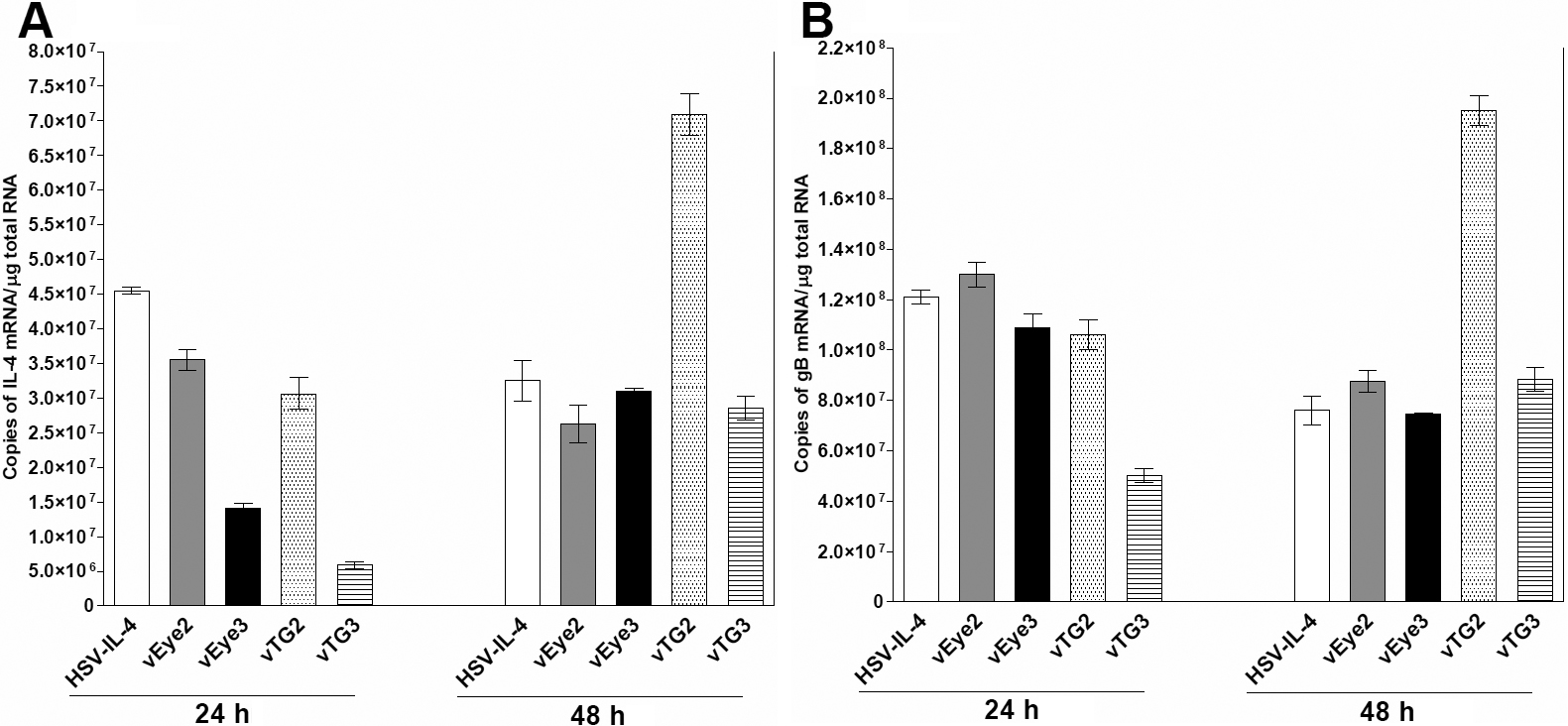
 Figure 6 of Mott, Mol Vis 2010; 16:2153-2162.