Figure 4. Digitalized images of
immunohistochemistry based protein expression, which were used for
quantification purposes. Increased TNF alpha expression was detected in
glaucomatous eyes (B), predominantly in the nerve fiber layer,
when compared to the control eyes (A). TNF alpha receptor 1
protein expression had similar appearance in control and glaucomatous
eyes (C, D). TNF alpha receptor 2 protein expression was
higher in glaucomatous eyes (F) when compared to control eyes (E).
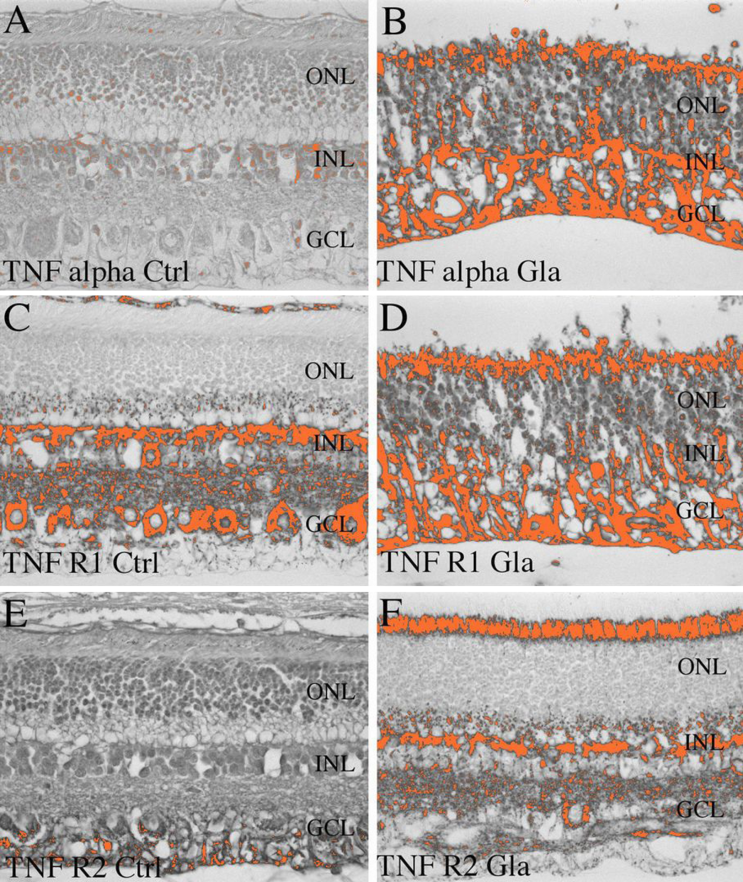
 Figure 4 of Jiang, Mol Vis 2010; 16:2092-2108.