Figure 5. Decreased inflammation in the
corneas of mice treated with pBTLA. A: pBTLA treated, pcDNA3.1
treated and PBS control mice were infected with 1×10⁵ PFU of
HSV-1 KOS. On day 14 postinfection, mice were euthanized, and eyes were
processed for cryosectioning. H&E staining was conducted on 5-µm
sections. The corneas in the pcDNA3.1 treated and PBS control groups
show severe swelling, heavy infiltration with inflammatory cells, and
numerous neovascular structures in the stroma. The cornea in the pBTLA
treated group exhibits only mild stromal edema, little inflammatory
cell infiltration, and fewer neovascular structures. Original
magnification, 400×. B: Six corneal samples from each group of
animals were digested with liberase on day 14 post infection. The bars
represent the total number of viable cells present per cornea of
different groups of mice. *p<0.01 compared with control groups. C:
The
cells isolated from infected corneas were stained for CD4 marker
and the bars represent the total number of CD4+ T cells
present per cornea from different groups of mice. Decreased numbers of
CD4+ T cells in the cornea of pBTLA treated mice. *p<0.01
compared with control groups. D: The percentage of CD4+
T cells in total cells of per cornea from different groups of mice.
*p<0.01 compared with control groups. E: Representative plot
shows CD4+ T cells expression on cornea of different group
mice.
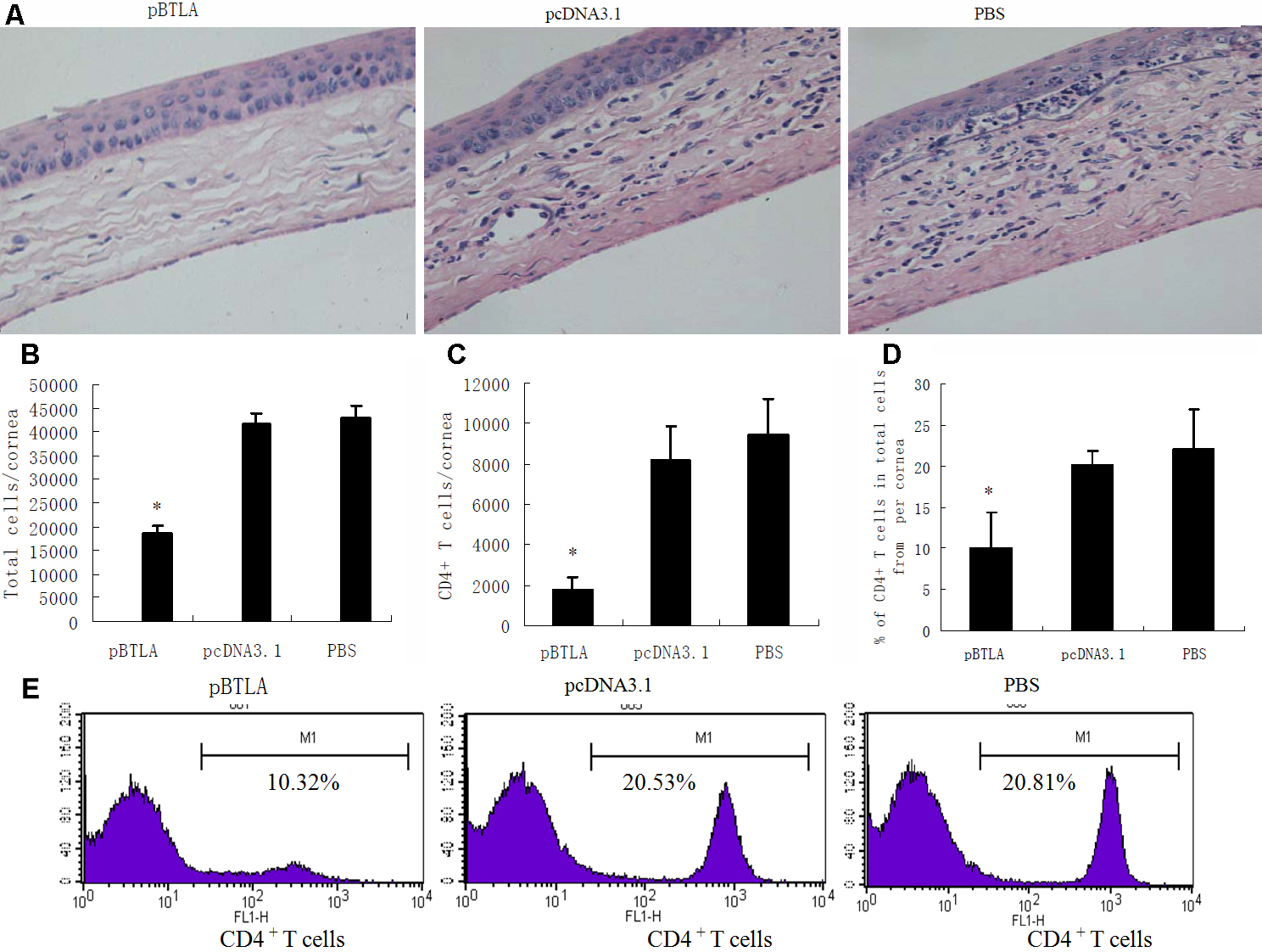
 Figure 5 of Xia, Mol Vis 2010; 16:2071-2083.