Figure 4. Treatment with pBTLA decreases the clinical severity of HSK. HSK lesion severity was determined by slit-lamp biomicroscopy,
and the clinical severity of stromal keratitis of individually scored mice was recorded. Stromal keratitis was graded from
0 to 4+, depending on the corneal opacity with neovascularization, edema, and infiltration. Each group of mice consisted of
fifteen animals, and the results shown are the representative of two similar experiments. A. Each dot represents corneal opacity
of an individual BALB/c eye on day 14 following HSV-1 infection. Crossbar indicates the mean. *p<0.01 versus control groups.
B. HSK lesion scores in three groups of mice at different time points post infection. *p<0.01 versus control groups.
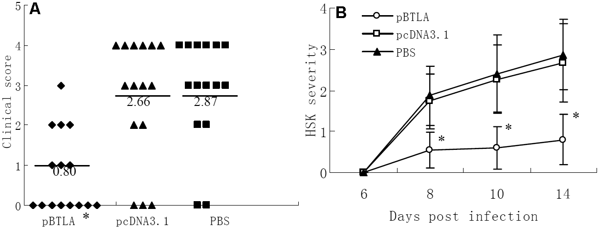
 Figure 4 of
Xia, Mol Vis 2010; 16:2071-2083.
Figure 4 of
Xia, Mol Vis 2010; 16:2071-2083.