Figure 3. Expression level of the recombinant plasmid pBTLA. A: Indirect immunofluorescence detection of BTLA in HEK293 cells. HEK293 cells were previously transfected with 1 μg of the
recombinant plasmids pBTLA or the empty vector, pcDNA3.1, in a 12-well plate. Forty-eight hours post-transfection, cells were
fixed with acetone, treated with anti-BTLA monoclonal antibody (mAb), and stained with FITC conjugated secondary antibody.
Green: BTLA protein-positive HEK293 cells transfected with pBTLA in fluorescence microscope. B: western blot result of recombinant plasmid pBTLA. HEK293 cells were transiently transfected with 1 μg of pBTLA. Cells were
harvested at 24, 48, 72 h after transfection, lysed, and the cell extract was resolved on a 12% SDS–PAGE gel. Protein was
transferred to a PVDF membrane (Bio-Rad) using a semi-dry transfer apparatus. Western blot membranes were scanned using an
Odyssey infrared imaging system (Trans-Blot SD, Bio-Rad). Lane 1 stands for the pcDNA3.1 transfected cell extracts, lanes
2–4 stand for the pBTLA transfected cell extracts at 24, 48, and 72 h post-transfection.
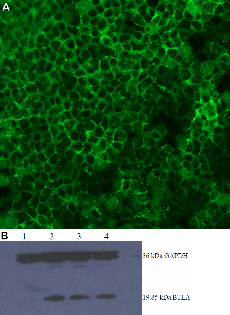
 Figure 3 of
Xia, Mol Vis 2010; 16:2071-2083.
Figure 3 of
Xia, Mol Vis 2010; 16:2071-2083.