Figure 1. BTLA expression is upregulated in murine peripheral blood and corneas after HSV-1 infection. The expression of BTLA on CD4+ and CD8+ T lymphocytes in murine peripheral blood was determined by flow cytometry on day 0, 3, 7, 10, 14, and 21 after corneal inoculation
with HSV-1 (n=6 at each time point). Peripheral blood mononuclear cells (PBMC) were stained simultaneously for CD3, CD4 or
CD8, and BTLA. PE mouse IgG1 was used as isotype control. A: The percentage of CD4+ T cells expressing BTLA at the indicated time point is shown in the bar diagrams. Statistically significant differences were
observed on day 3, 7, 10, and 14 compared to day 0 (*p<0.01). B: Representative three-color flow cytometry plot of pre-infection (day 0) versus day 10 post-infection, gated on CD4+ T cells indicating BTLA expression in murine peripheral blood. C: The percentage of CD8+ T cells expressing BTLA for the indicated time point is shown in the bar diagrams. No statistically differences were observed
on day 3, 7, 10, 14 and 21 compared to day 0 (p>0.05). Corneal tissues (D,E) were analyzed by immunohistochemistry to determine BTLA expression. Immunohistochemical staining with an antibody against
BTLA was performed to detect BTLA expression. Sections incubated without a primary antibody served as negative controls. All
tissue sections were counterstained with hematoxylin. Brown staining indicates BTLA expression. D: Corneal tissues derived from a normal uninfected mouse. Faint BTLA expression was only observed in the corneal epithelium
(arrow). E: Cornea tissue derived from a mouse 10 days after corneal HSV-1 inoculation. Increased BTLA expression was observed in the
corneal epithelium, stroma, endothelium, and many inflammatory cells that infiltrated the corneal stroma (arrows). Original
magnifications, 400×.
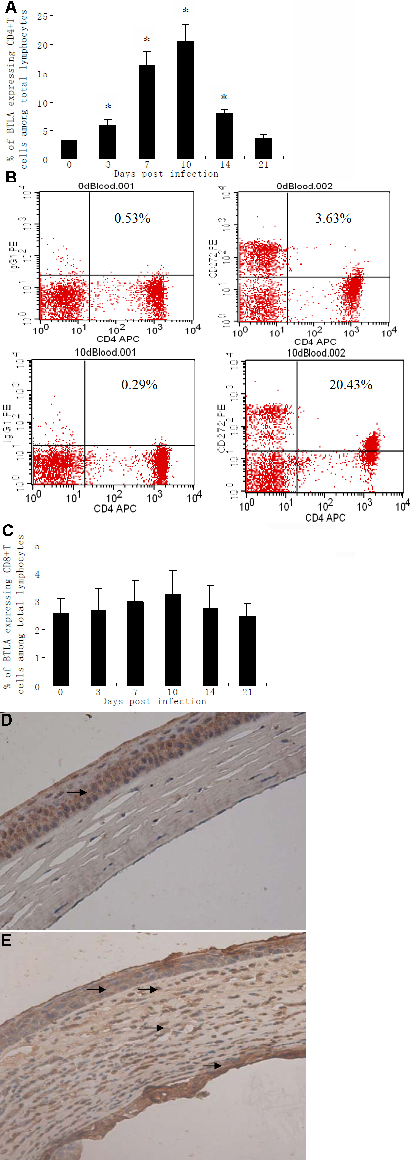
 Figure 1 of
Xia, Mol Vis 2010; 16:2071-2083.
Figure 1 of
Xia, Mol Vis 2010; 16:2071-2083.