Figure 5. H-RN inhibits retinal
neovascularization. A: Retinal vascular distribution of a
normal pup raised in room air. B: This figure shows a
representative retina from 17-day-old pups that were subjected to 5
days of 75% oxygen tension and then maintained in room air for an
additional 5 days. Circles indicate the neovascular tufts. C: A
retina from a mouse pup injected with vascular endothelial growth
factor ab (VEGFab; 50 ng/µl). D: A retina from a mouse pup
injected with H-RN (50 mM). E: A retina from a mouse pup
injected with H-FT (50 mM). F: A retina from a mouse pup
injected with scrambled peptide (50 mM, magnification, ×40). G:
A hematoxylin- and eosin-stained section of P17 control raised in room
air. H: P17 retina exposed to hyperoxia from P7 to P12, and
returned to room air from P12 to P17. Arrows indicate neovascular tufts
extending into the vitreous. I: A section from 50 ng/µl
VEGFab-treated hyperoxia group. J: A section from 50 mM
H-RN-treated hyperoxia group. The retina of I and J
presented obvious reduced abnormal neovascularization tufts (arrows). K:
A
section from 50 mM H-FT-treated hyperoxia group. L: A section
from 50 mM scrambled peptide treated hyperoxia group (magnification,
×400). M: Quantification of neovascular lumens response to
hyperoxia. Lumens extending from the internal limiting membrane into
the vitreous were counted. Data in each column present means±SD values
of total number of vascular lumens per retinal cross-section, from 7 to
9 eyes of 7–9 mice. Note that the lumens in VEGFab and H-RN-treated
group are reduced significantly more than the control and PBS groups
(p<0.001), while the number of lumens in the H-FT and scrambled
peptide-treated group had no difference from the control and PBS
treated group (p>0.05). These experiments were repeated three times
with similar results (** p<0.01, each condition versus control).
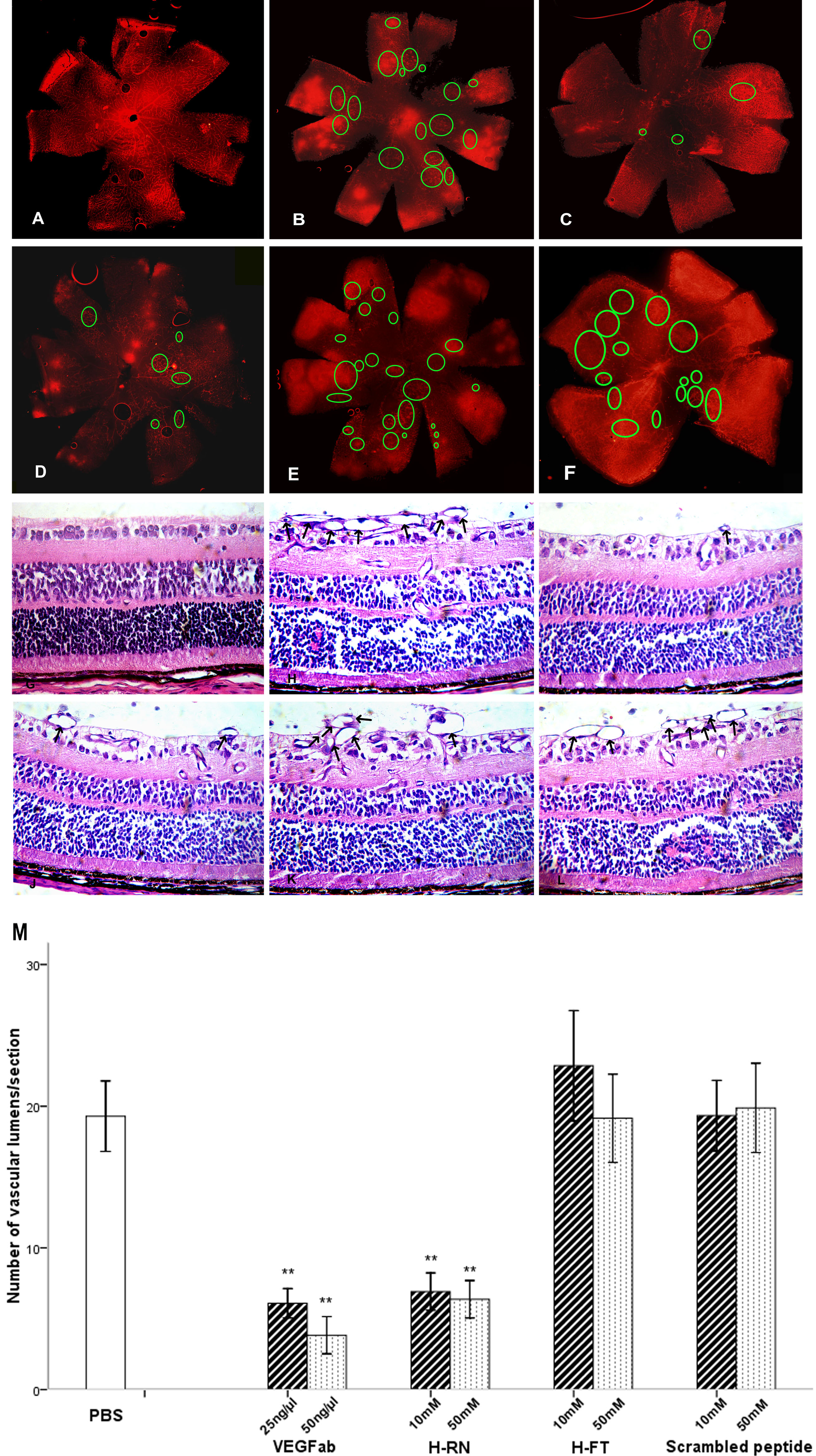
 Figure 5 of Xu, Mol Vis 2010; 16:1982-1995.