Figure 3. Inhibition effects of H-RN and
H-FT on endothelial cell differentiation into capillary structures.
Starved RF/6A cells were pre-incubated with different concentrations of
bevacizumab, H-RN, and H-FT for 30 min before 100 ng/ml vascular
endothelial growth factor (VEGF) was added in peptides treated and VEGF
groups, and then cells were seeded into Matrigel-coated wells. A-F:
Representative
photographs of anti-tube formation activity of
bevacizumab, H-RN, H-FT, and scrambled peptide: A, no-VEGF; B,
100
ng/ml VEGF; C, 100 ng/ml VEGF +2.5 mg/ml bevacizumab; D,
100
ng/ml VEGF +1 mM H-RN; E, 100 ng/ml VEGF +100 nM H-FT; F,
100
ng/ml VEGF +1 mM scrambled peptide. VEGF at 100 ng/ml strongly
stimulated endothelium tube formation. Bevacizumab and H-RN potently
inhibited VEGF-stimulated RF/6A cell tube formation on growth
factor-reduced Matrigel™. H-FT and scrambled peptide did not inhibit
RF/6A cell tube formation effectively (magnification ×200). G:
Quantitative analysis of tube formation under the different
experimental conditions using Image Program Plus software. The values
are mean tube lengths from three repeated experiments (** p<0.01,
each condition versus control).
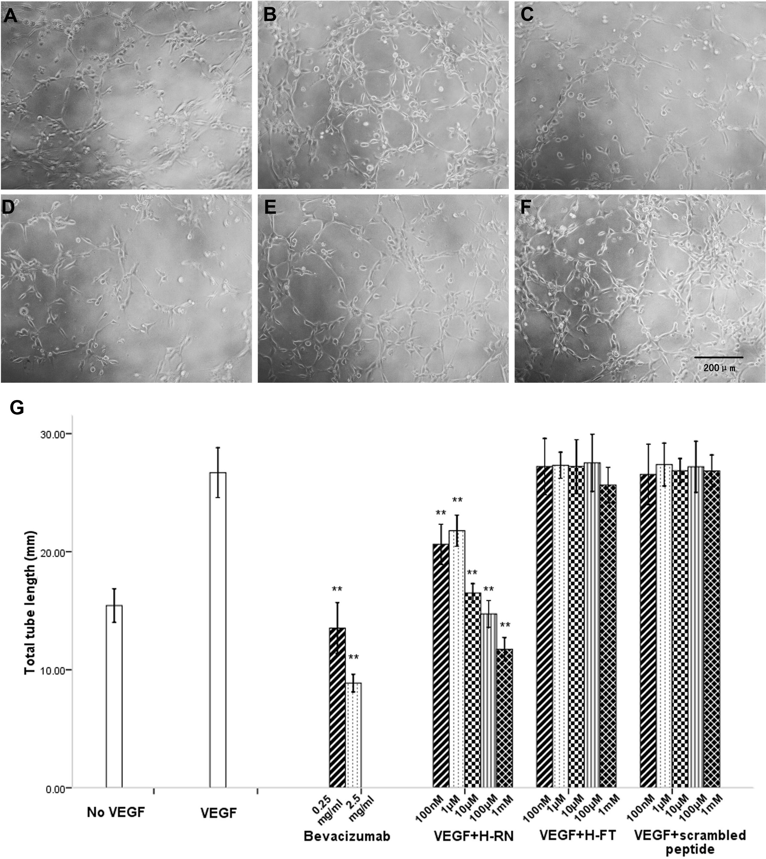
 Figure 3 of Xu, Mol Vis 2010; 16:1982-1995.