Figure 6. Inflammatory changes in
recoverin-immunized B6.MRL-Fallpr/J (LPR) mice
and hybridoma balb/cJ mice. Increase of C1q and C3 deposition and
infiltration of CD3+ and CD68+ cells in the retina of
recoverin-immunized LPR mice and Balb/c mice injected
with recoverin hybridoma cells. No C1q deposition and C3 deposition in
only retinal pigment epithelium (RPE) was observed in control LPR mice,
while they were enhanced in the RPE, inner nuclear layer (INL), and
ganglion cell layer (GCL) of recoverin-immunized LPR mice retina. No
C1q or C3 deposition in RPE was observed in control Balb/c
mice, while some C1q deposition was observed in the RPE, INL and GCL of
the retina of Balb/c mice injected with recoverin hybridoma
cells, and a marked increase of C3 deposit on RPE and INL was observed.
CD3+ cells were observed between the INL and GCL layers, and the
migrating cells between INL and GCL were CD3+ cells. While CD68+ cells
were found in inner plexiform layer (IPL) and outer plexiform layer
(OPL) in recoverin-immunized LPR mice retina, they were found in RPE
and OPL in Balb/c mice injected with recoverin hybridoma cells.
Cell nuclei were stained with 4',6-diamidino-2-phenylindole (DAPI;
blue). Abbreviations: ONL, outer nuclear layer. Scale equal to 50 µm.
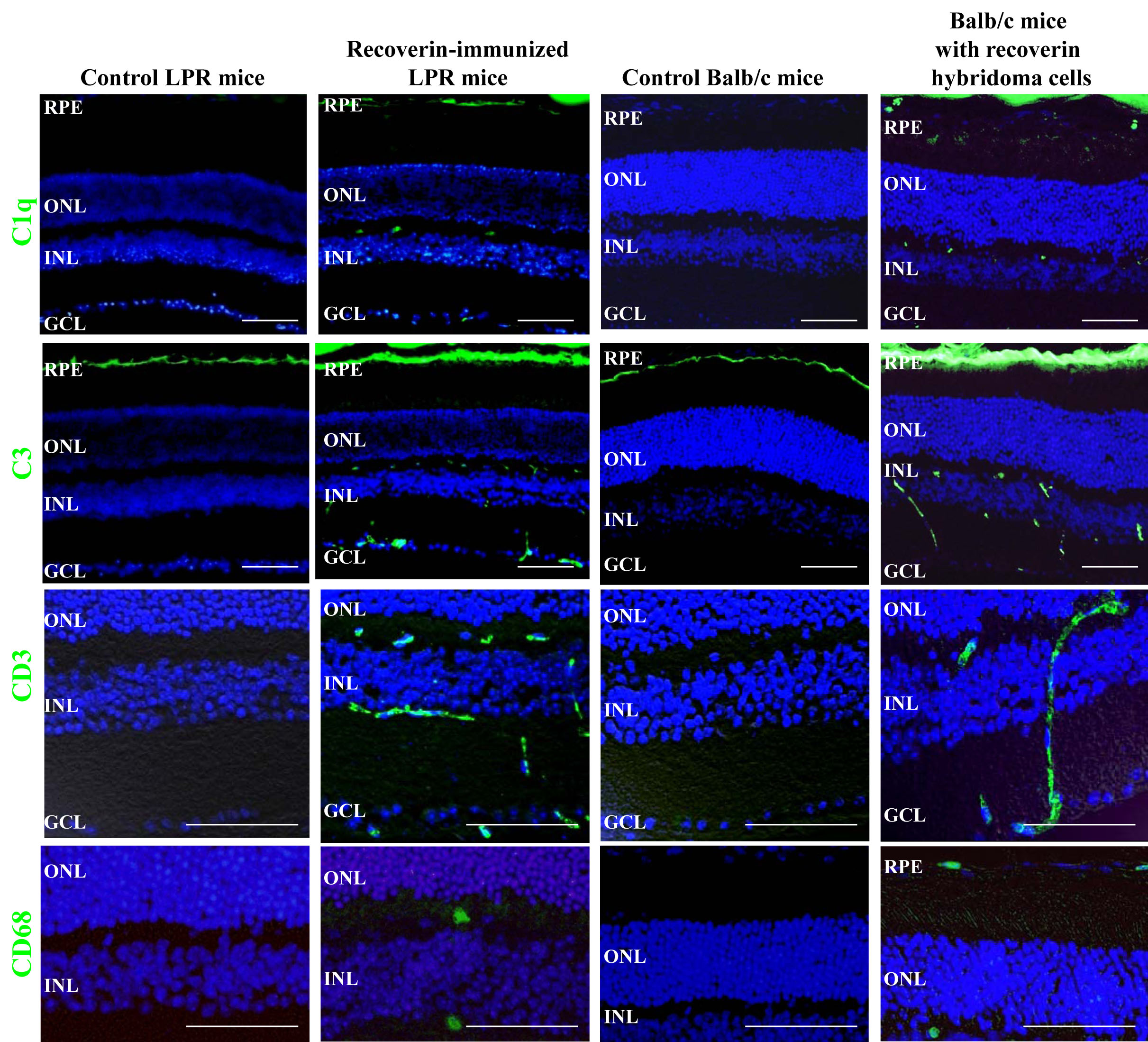
 Figure 6 of Lu, Mol Vis 2010; 16:1936-1948.