Figure 4. Analysis of innate immune cells
into a corneal graft. Ox-62+ DC and CD163+
macrophages were stained at the time points of corneal allograft
rejection and calculated within the graft. Additionally CD161+
cells were counted within rejected corneal allografts to finally prove
the efficacy of the depletion protocol in the peripheral tissue.
Representative histological staining is shown for Ox-62 (A),
CD163 (C), and CD161 (E) in NK deficient and control
animals. B: No statistical difference was observed for
infiltrating Ox-62+ DC. D: CD163+
macrophages infiltrated to a statistically significantly stronger
extent in 3.2.3-treated animals when compared to control treated
animals (*p<0.01). F: No CD161+ cells were
stained in 3.2.3-treated recipients when compared to control treated
control animals (**p<0.001).
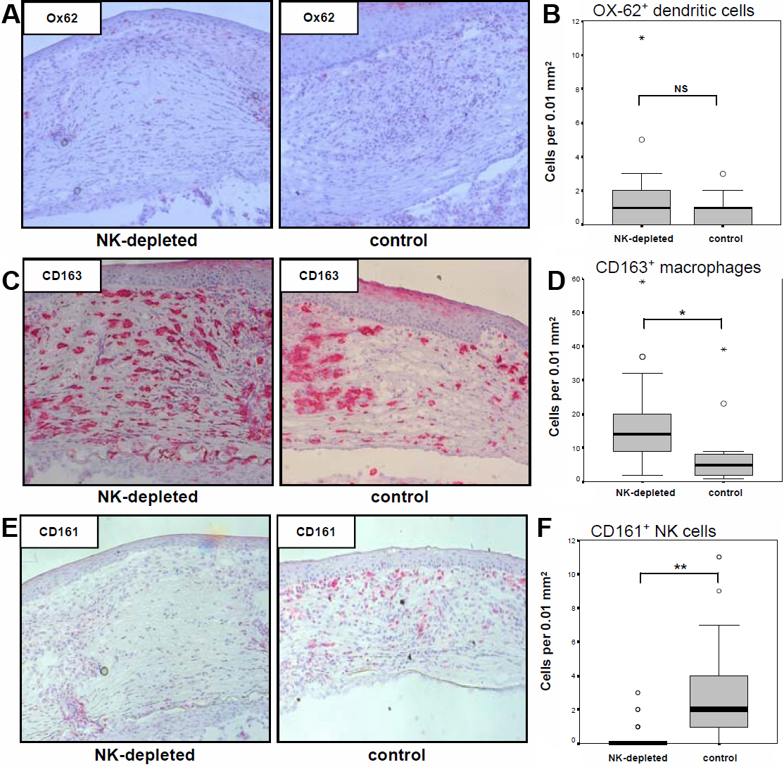
 Figure 4 of Schwartzkopff, Mol Vis 2010; 16:1928-1935.