Figure 2. Analysis of graft-infiltrating
leukocytes. At the time points of corneal allograft rejection CD45+
leukocytes were stained and calculated within the graft. A:
Histological staining of graft infiltrating CD45+ leukocytes
is shown exemplarily. B: Calculation from 4 representative
grafts revealed no statistically significant differences in the total
amount of infiltrating cells in rejected grafts of 3.2.3- and control
treated recipients, respectively (NS=not significant).
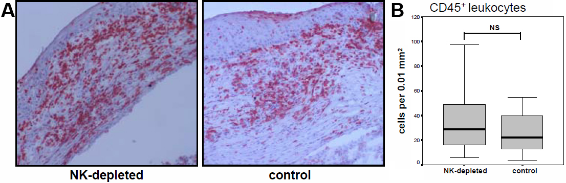
 Figure 2 of Schwartzkopff, Mol Vis 2010; 16:1928-1935.