Figure 1 of
He, Mol Vis 2010; 16:1913-1919.
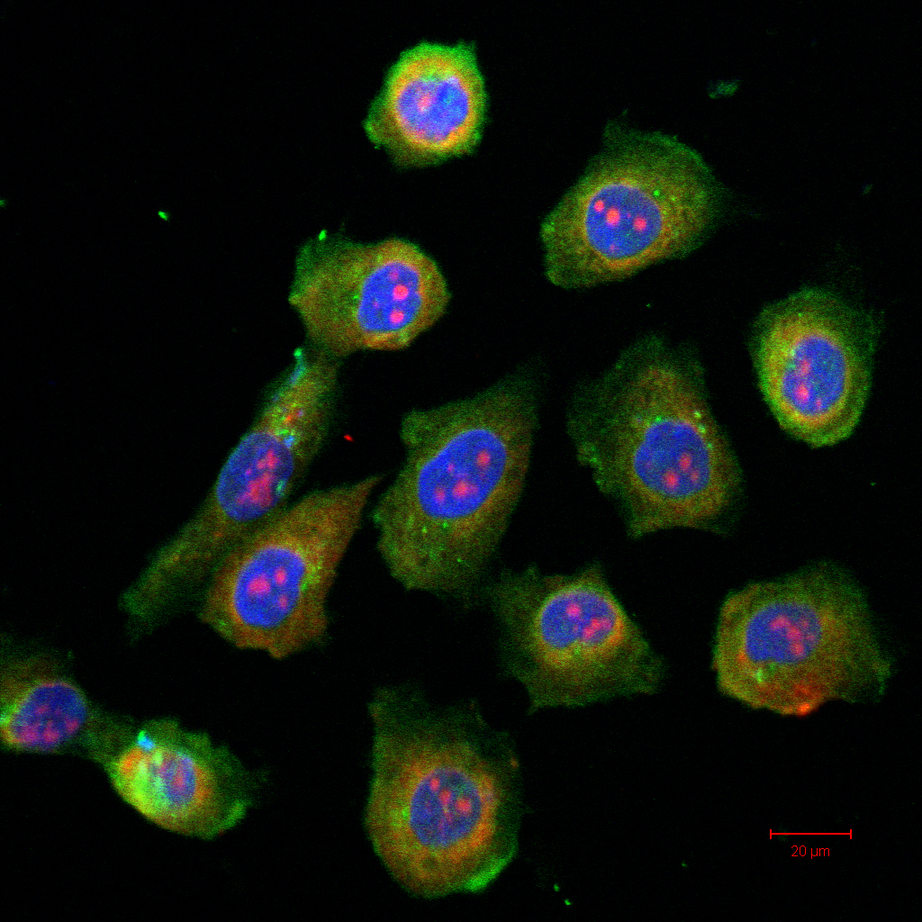
Figure 1.
Photomicrographs of CGCs in vitro. Double immunolabeling for CK-7 and MUC5AC indicate the presence of CK-7 (shown in green) and MUC5AC (shown in red) in the same cell in culture. The scale bar represents 20 μm.
 Figure 1 of
He, Mol Vis 2010; 16:1913-1919.
Figure 1 of
He, Mol Vis 2010; 16:1913-1919.