Figure 3. Exon 16 splicing pattern
associated with the c.1935G>A mutation. A: Analysis of
endogenous RNA from lymphoid cells. We amplified a 346-bp fragment from
exon 14 to exon 17 of MYO7A in a healthy control individual. An
additional faint band appeared in the control; it corresponded to an
mRNA species missing exon 16 (lane 2). In patient MB9, the splicing
pattern showed a predominant exclusion of exon 16 (lane 1). Lane 3
refers to negative control and M indicates size marker. Direct
sequencing of these two different transcripts revealed that a shorter
band of 246 bp corresponds to an abnormal transcript without exon 16,
whereas the other band of 374 bp corresponds to a normal transcript
that contains exon 16. B: Ex vivo splicing assays were
performed. Wild-type and mutant constructs were stably transfected in
HeLa cells, and the exon 16 splicing pattern was analyzed by reverse
transcription-polymerase chain reaction. Consistent with data presented
in A, exon 16 is predominantly included from the wild-type construct
(lane 1). The c.1935G>A mutation at the end of exon 16 resulted in
massive skipping of the exon (lane 2). Lane 3 refers to negative
control and M indicates size marker. UE and DE refer to upstream and
downstream exons of the cassette, respectively.
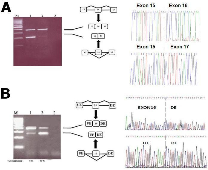
 Figure 3 of Ben Rebeh, Mol Vis 2010; 16:1898-1906.