Figure 9. Immunofluorescence for CTGF and α-SMA in tissue sections from eyes five days after glaucoma filtration surgery (GFS). The
top row; immunofluorescence staining for CTGF (red), the bottom row; immunofluorescence for α-SMA (green). Diffuse subconjunctival
cell staining for both CTGF and α-SMA was observed in the no adjunct control while weaker and more scarce staining was demonstrated in the SB-505124 and MMC
groups. The weaker and less staining seems due to suppression of CTGF/α-SMA expression in the SB-505124 group. In the MMC group, it is based on a lower cell number. Arrows indicate the conjunctival
epithelium; asterisk, the subconjunctival space; S, the sclera; arrowhead, blood vessel with red blood cells; and double arrowhead,
non specific staining. Bar, 100 μm.
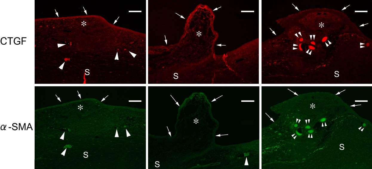
 Figure 9 of
Sapitro, Mol Vis 2010; 16:1880-1892.
Figure 9 of
Sapitro, Mol Vis 2010; 16:1880-1892.