Figure 8. Hematoxylin and eosin staining in tissue sections from eyes five days after surgery. Infiltration of only a few inflammatory
cells and mild scarring were observed in the subconjunctival space of eyes in the GFS with SB-505124 (left panel) or the MMC
control (center panel) group, whereas numerous inflammatory cells and massive scarring were seen in the no adjunct controls
(right panel). At the corneal limbus, infiltration of cells was observed in all 3 groups. Thinner conjunctival epithelium
was seen in the MMC control compared to that in the GFS with SB-505124 and no adjunct control groups. Subconjunctival blood
vessels were noted in GFS with SB-505124 and no adjunct control groups in general, but seldom in the MMC control group. Arrows
indicate the conjunctival epithelium; double arrow, the limbus; arrowhead, the subconjunctival blood vessel; asterisk, the
subconjunctival space; and S, the sclera. Bar, 200 μm.
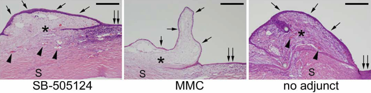
 Figure 8 of
Sapitro, Mol Vis 2010; 16:1880-1892.
Figure 8 of
Sapitro, Mol Vis 2010; 16:1880-1892.