Figure 4. Immunofluorescence staining for CTGF and α-SMA in subconjunctival fibroblasts. Immunoreactive products were visualized with
Alexa Fluor (green) labeling. Nuclei were stained with DAPI (blue). Rabbit subconjunctival fibroblasts were treated with or
without TGF-β2 and/or SB-505124. A dramatic increase in staining for TGF- β2-induced proteins CTGF and α-SMA was observed
following TGF-β2 incubation. The staining intensity of these proteins was markedly reduced when cells were treated concomitantly
with SB-505124. The cell density was not affected by SB-505124 treatment. Bar, 50 µm.
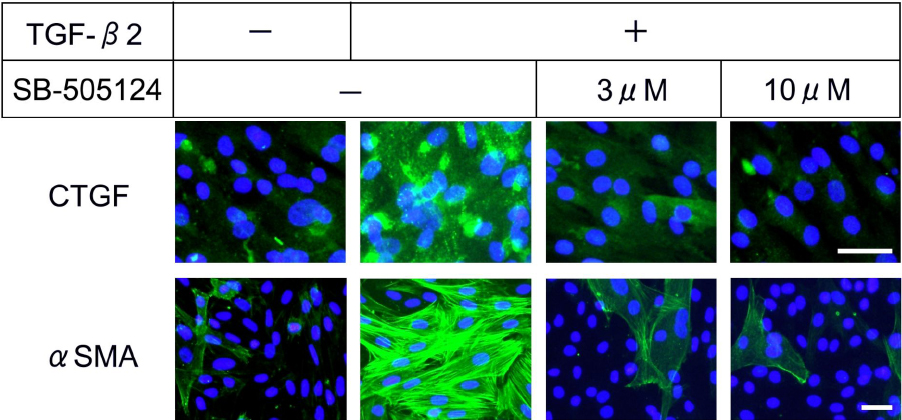
 Figure 4 of
Sapitro, Mol Vis 2010; 16:1880-1892.
Figure 4 of
Sapitro, Mol Vis 2010; 16:1880-1892.