Figure 3. Western blotting for phosphorylated Smad2 (pSmad2), CTGF, and α-SMA. Rabbit subconjunctival fibroblasts were incubated with
TGF-β2 and various concentrations of SB-505124. SB-505124 effectively reduced the pSmad2 level and the expression of CTGF
and α-SMA induced by TGF-β2 in a concentration-dependent fashion. SB-505124 was effective when its concentration reached 1
µM, and the expression of CTGF and α-SMA at 3 and 10 μM of concentrations is below that in cells without the TGF-β2 stimulation.
The blots were also probed for Smad2, β-actin, or GAPDH. The band intensity of pSmad2, CTGF and α-SMA was normalized against
that of Smad2, β-actin, and GAPDH, respectively, and the ratios are shown.
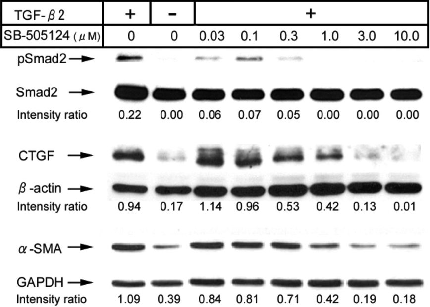
 Figure 3 of
Sapitro, Mol Vis 2010; 16:1880-1892.
Figure 3 of
Sapitro, Mol Vis 2010; 16:1880-1892.