Figure 10. Fibroblast outgrowth from subconjunctival tissues. Five days after GFS, subconjunctival tissues were dissected, and each biopsy
specimen was placed in a 25 cm2 cell culture flask with complete media. Image of fibroblast outgrowth from subconjunctival tissues at day 10 in culture is
shown. Cell outgrowth from explants of the GFS with SB-505124 (left panel) and the 180° control (right panel) groups is robust
whereas outgrowth is poor from that of the MMC control group (center panel). A few small, round specks that appeared to be
dead cells were found on top of the outgrowth from tissue treated with inhibitor and MMC, but seldom in that from the 180°
control tissues. This suggests that, while its toxicity is vastly reduced compared to MMC, 5 mg of SB-505124 delivered within
a lactose tablet is not entirely nontoxic.
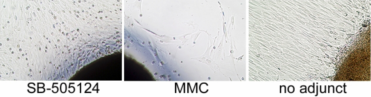
 Figure 10 of
Sapitro, Mol Vis 2010; 16:1880-1892.
Figure 10 of
Sapitro, Mol Vis 2010; 16:1880-1892.