Figure 2. Co-localization of Caspr and inl
cell markers in rodent retina. A-C: Single confocal
section demonstrating co-localization of Caspr labeled amacrine cells
near the inner nuclear layer/inner plexiform layer (inl/ipl) boundary (A,
arrowheads)
with
Disabled-1
(B) a marker of AII amacrine cells
in mouse retina (C, overlay) D-F:
Photomicrographs of rat retina demonstrating that nearly all Caspr
labeled amacrine cells (e.g., arrowheads, D) also contained
Parvalbumin (PV, E), a well known marker for AII amacrine cells
(F, overlay). Arrow in (D) indicates a cell labeled with
Caspr, but not co-localized with Parvalbumin. Asterisk in (E)
indicates a cell labeled with Parvalbumin, but not co-localized with
Caspr. G-I: Photomicrographs of rat retina demonstrating that
Caspr (G) was not co-localized with ChAT (H) in amacrine
cells (I, overlay). Arrowheads indicate Caspr labeled cells in
the inl; arrows indicate ChAT labeled cells in the GCL. J-L:
Photomicrographs of rat retina showing that some Caspr-labeled amacrine
cells (arrowheads, J) contained Calretinin (K
arrowheads, L overlay). Arrows in J-L indicate a
Caspr labeled amacrine cell that did not co-localize with Calretinin,
while asterisks (J-L) indicate a Calretinin positive cell
that did not contain Caspr. Scale bar equal to 25 μm.
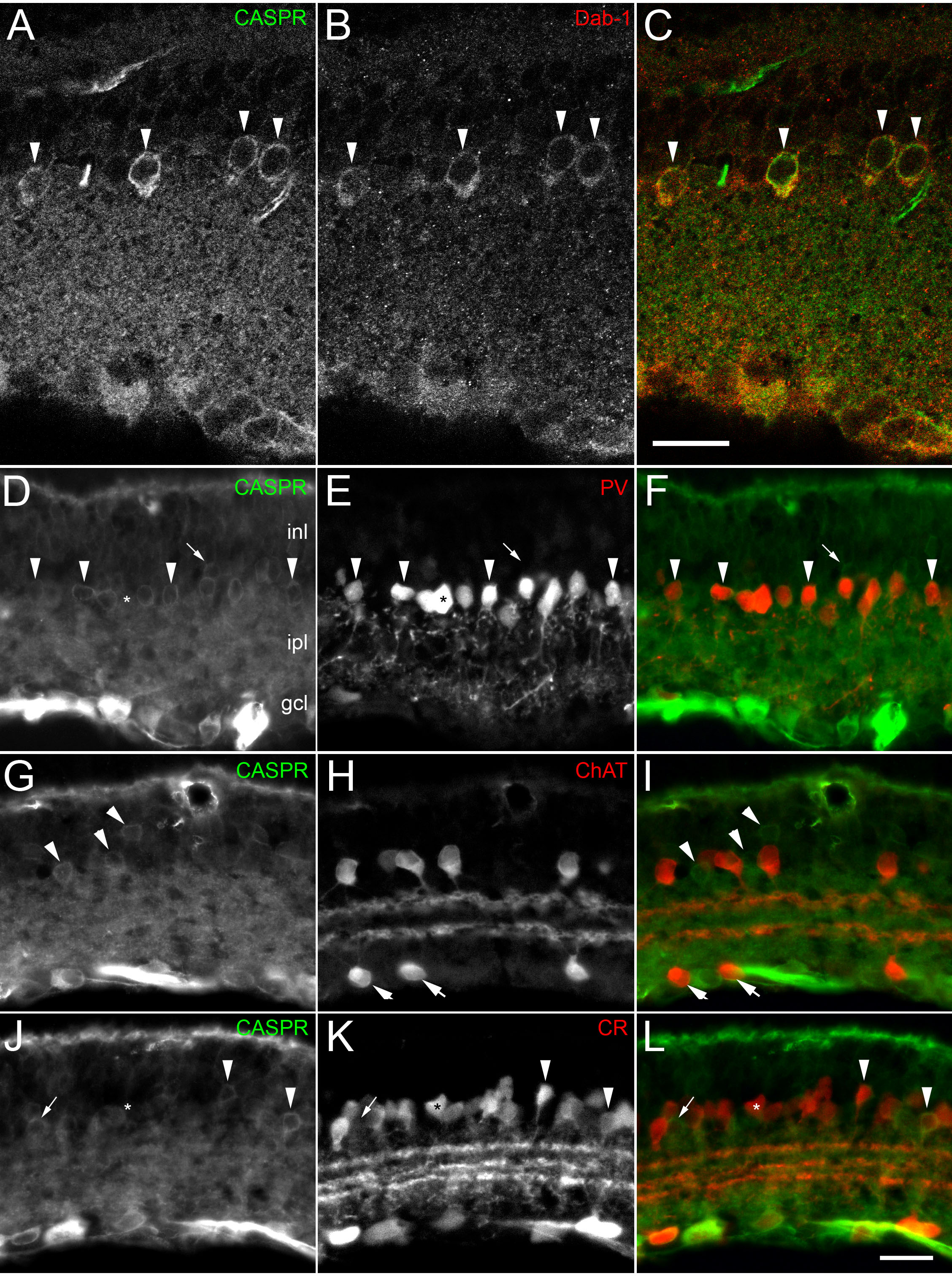
 Figure 2 of O’Brien, Mol Vis 2010; 16:1854-1863.