Figure 1. Caspr labeling in rat and mouse
retina. A: Photomicrograph of monoclonal Caspr labeling in rat
retina. Arrow indicates one of several retinal ganglion cells (RGCs)
intensely labeled by Caspr. In addition to RGCs, somas of many amacrine
cells in the inner nuclear layer (inl) were also labeled (e.g.,
arrowheads). The inner plexiform layer (ipl) was diffusely labeled
while the outer plexiform layer (opl) contained several hot spots. B:
Lower
power
photomicrograph
of Caspr labeling in rat retina using a
rabbit polyclonal antibody. A nearly identical pattern of labeling was
observed as in A. C: Single confocal section of rat
retinal wholemount labeled with a monoclonal Caspr antibody. Intense
labeling of axon fiber bundles was observed (arrowheads) as well as
somas of nearly all cells in the ganglion cell layer. Both large RGCs
(asterisk) and likely displaced amacrine cells (arrow, somas <10 μm)
were labeled. D: Photomicrograph of Caspr labeling in the rat
optic nerve. Inset shows magnified view of node indicated. Arrow
indicates another labeled node. E: Photomicrograph of Caspr
labeling (mAb) in mouse retina. Similar to the rat retina (A, B)
intense
labeling
of
RGCs and fiber bundles were observed as well as
many amacrine cells in the inl. F: Photomicrograph of Caspr
labeling (mAb) in knockout retina. All labeling of retinal cell types
was eliminated, leaving only nonspecific labeling of retinal blood
vessels. Scale bars equal to 25 μm.
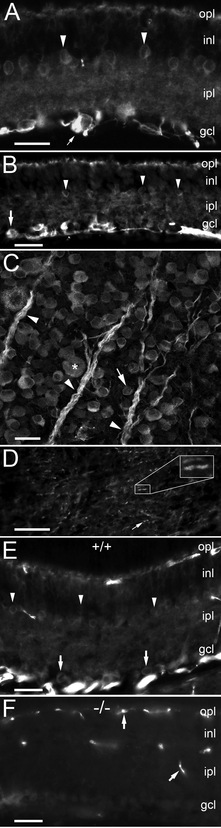
 Figure 1 of O’Brien, Mol Vis 2010; 16:1854-1863.