Figure 2. Effects of vascular endothelial
growth factor (VEGF) in oxidative stress induced cytotoxicity in
retinal ganglion cell (RGC)-5 cells. A: Hydrogen p 258 eroxide
dose response curve, following 24 h exposure, revealed 30%–35%
reduction in cell numbers at 700 and 800 μM doses. Higher doses
resulted in 80% reduction (*p-value<0.005). B: VEGF-mediated
protection was evaluated in parallel wells, where hydrogen peroxide
(800 μM)-treated cells are compared with cells treated with VEGF alone.
There was a reduction in percentage decrease of cell numbers when cells
were co-treated with VEGF and H2O2, compared with
controls administered in a dose-dependent manner with increasing doses
of VEGF. The maximum protection was provided at the 200 nM dose
(p=0.001). C: Treatment with the glutathione reductase
inhibitor buthionine sulfoxime (BSO) increased H2O2-mediated
reduction
in cell numbers by 22%. hVEGF165 at 200 ng/ml provided the
greatest protection, increasing cell numbers by 8%, p<0.0005. Cell
numbers were determined using a Water Soluble Tetrazolium (WST)-1 assay
and are expressed as percent of control. H2O2:
hydrogen peroxide.
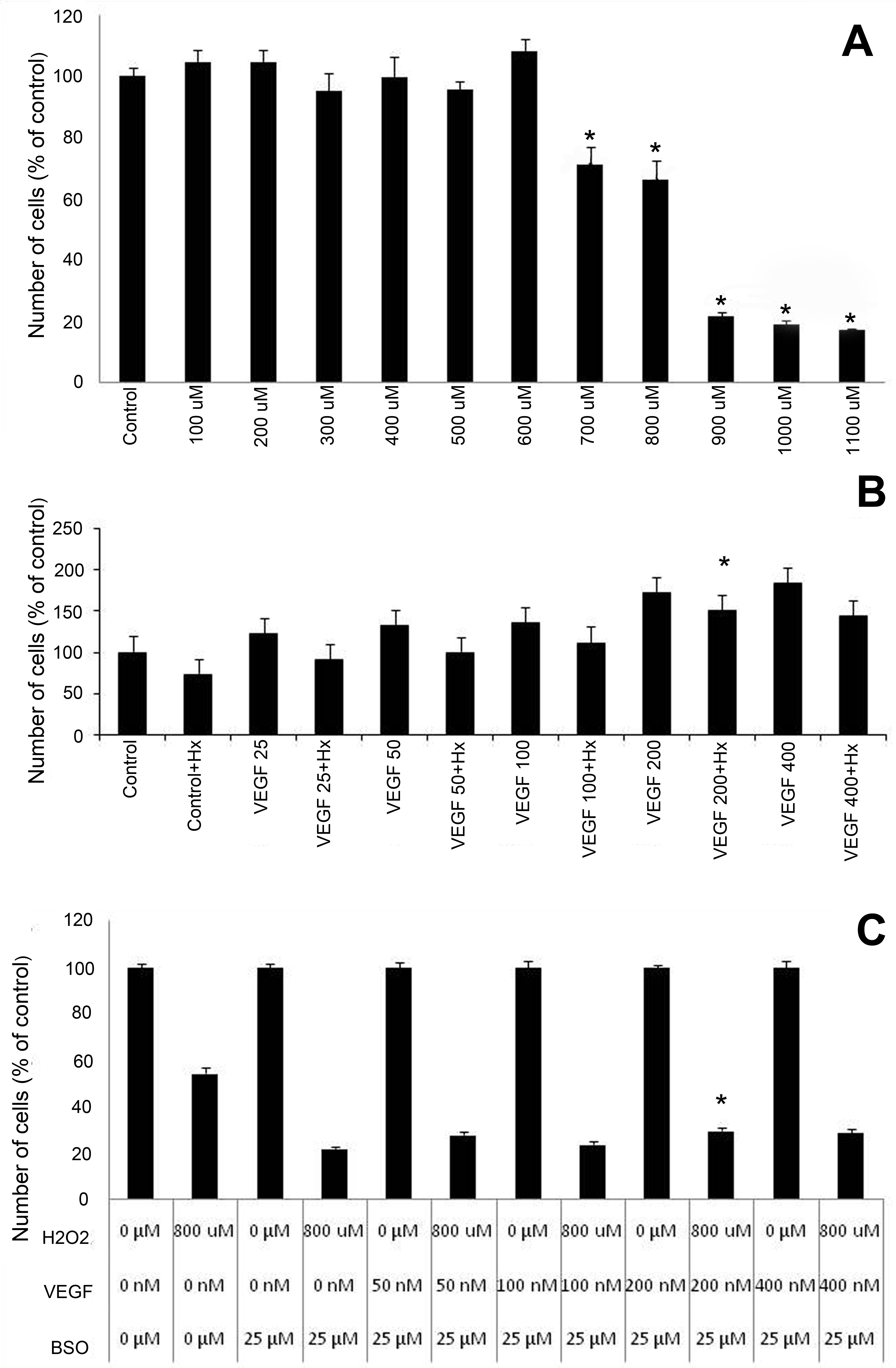
 Figure 2 of Brar, Mol Vis 2010; 16:1848-1853.