Figure 1. Morphological and
immunohistochemical differentiation of retinal ganglion cell (RGC)-5
cells. A: Phase contrast biomicroscopy comparing
undifferentiated with differentiated RGC-5 following exposure to
Staurosporine. Note the elongated neuronal processes in the treated
cells. B: Antibody control for immunocytochemistry staining C:
Immunocytochemistry
staining for class III β-tubulin highlights the
elongated processes in Staurosporine treated cells. undiff:
undifferentiated, diff: differentiated, AB: antibody control.
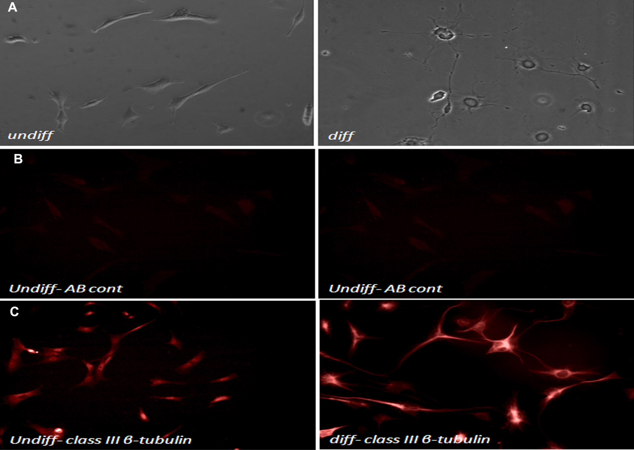
 Figure 1 of Brar, Mol Vis 2010; 16:1848-1853.