Figure 1. Retinal dysplasia and degeneration (rdd) retinal histology. Microtome sections of retina stained with hematoxylin and eosin. Normal retinal morphology is evident
in the developing wt chick at embryonic day (E)13 (A), E18 (C), and post-hatch day (P)1 (E). At E13 the gross retinal morphology of the rdd retina (B) is similar to that of the wt retina; however, the degenerative changes are obvious in the rdd retina at E18 (D) and P1 (F). Magnified images of the outer plexiform layer (OPL) and outer nuclear layer (ONL) show the progressive disorganization
of the outer retinal layers of the rdd chick from E13 (I), E18 (J), and P1 (K).
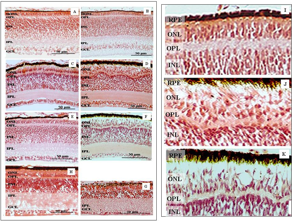
 Figure 1 of
Finnegan, Mol Vis 2010; 16:7-17.
Figure 1 of
Finnegan, Mol Vis 2010; 16:7-17.