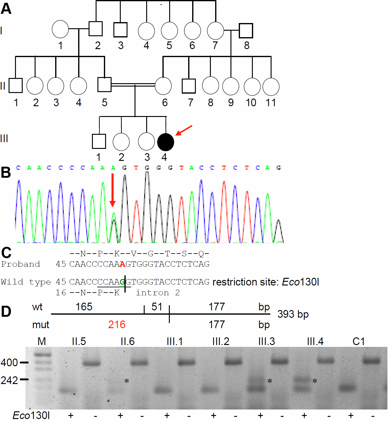
Figure 5. CRYBB2 mutation in family DJC1.
A: The pedigree of family DJC1 indicates that a zonular cataract appeared in the youngest daughter of healthy, but consanguineous
parents. There is no other report of any type of cataract over three generations.
B: Sequence analysis of
CRYBB2 genomic DNA indicates heterozygosity for the proband at cDNA pos. 54 (red arrow).
C: Comparison of the wild-type sequence (
Z99916) with the proband’s sequence demonstrates that the G→A exchange at cDNA-position 54 leads to an altered splice site; it is
predicted that the mutated mRNA contains at least part of intron 2 (the A of the ATG start codon is counted as #1; in the
amino-acid sequence, the first Met is counted as #1). The mutation leads to a loss of an Eco130I restriction site.
D: Restriction analysis using the enzyme Eco130I creates a larger fragment of 216 bp in the mutant DNA (red); it demonstrates
the presence of the mutation in the proband (III.4), but surprisingly also in the healthy mother (II.6) and one healthy sister
(III.3) of the proband; the asterisks mark the additional band of 216 bp indicating the mutation. Undigested PCR fragments
of 393 bp are indicated by “-” or “+” are digested PCR products. The small fragment of 51 bp in the wild-type DNA is not visible;
the two bands of 165 and 177 in the wild type are not separated under the conditions used here. C1 and C2 represent independent
controls from the laboratory; M=marker.
 Figure 5 of
Santhiya, Mol Vis 2010; 16:1837-1847.
Figure 5 of
Santhiya, Mol Vis 2010; 16:1837-1847.