Figure 2. CRYGD mutation in the family CBE21. A: Sequence analysis of exon 2 of CRYGD indicates heterozygosity (arrow) for the mother of the proband (III.4). B: Comparison of the wild-type sequence (WT) with the proband’s mother’s sequence (CBE21) demonstrates that the C→T mutation
at cDNA position 130 leads to an amino acid exchange from Met to Val at pos. 44 (M44V); moreover, it shows the creation of
a new Alw21I restriction site (underlined) in two (III.4 and III.6) of the affected members of the family checked, while the
same was absent in the proband’s grand mother (II.2). One of the proband’s uncle (III.5) could not be checked (in the cDNA
sequence, the A of the ATG start codon is counted as #1; in the amino-acid sequence, the first Met is counted as #1). C: Restriction analysis with (+) or without (-) the enzyme Alw21I in the members of the core family leads to an additional
fragment of 370 bp in the mutant DNA (red); it demonstrates the presence of the mutation in the affected mother (III.4) and
the affected brother of the proband’s mother (III.6). The asterisks mark the additional band of 370 bp indicating the mutation;
the band of 533 bp indicates the undigested DNA. WT represents an independent control from the laboratory; M=marker.
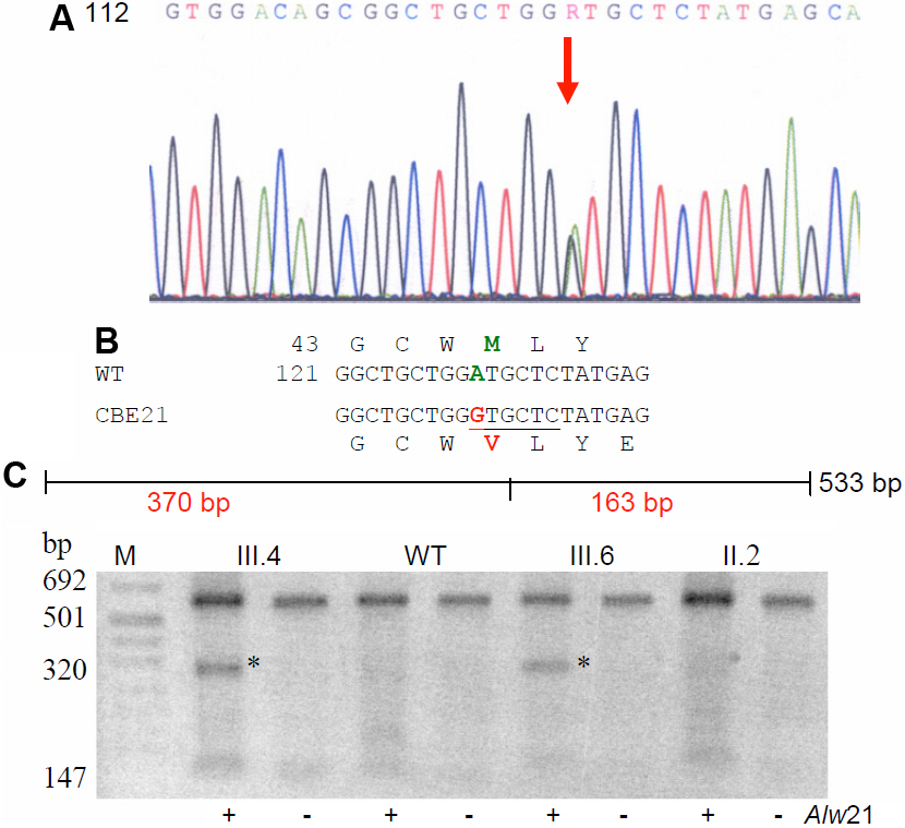
 Figure 2 of
Santhiya, Mol Vis 2010; 16:1837-1847.
Figure 2 of
Santhiya, Mol Vis 2010; 16:1837-1847.