Figure 8. Effect of elevated cAMP on the
organization of microtubules and PAMR. Cells were treated with 20 ng/ml
TNF-α (T) for 6 h and where indicated were co-treated with 10 µM
forskolin (F) and 50 µM rolipram (R). A-D: Organization
of microtubules. In untreated cells (Control, A), microtubules
exist as characteristic fibrillary extensions from around the nucleus
to the cell periphery (shown by arrows). It is undisturbed by F and R (B).
Treatment
with
TNF-α (C) induced microtubule disassembly
characterized by the loss of as well as condensation of fibrillary
extensions (shown by arrows), which was opposed by co-treatment with F
and R (D). E-H: Organization of PAMR. In
untreated cells (Control, E), the characteristic organization
of cortical actin with intact PAMR is observed (shown by arrows). It is
undisturbed by F and R (F). Treatment with TNF-α induced
disruption of PAMR (G) (shown by arrows), which was opposed by
co-treatment with F and R (H). All the images shown are
representative of at least three independent experiments.
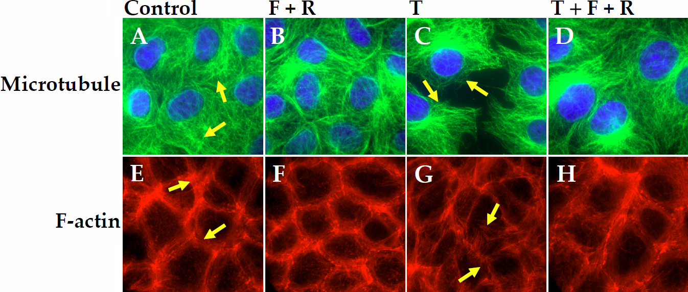
 Figure 8 of Shivanna, Mol Vis 2010; 16:1781-1790.