Figure 6. Immunohistochemical localization
of OGG1 and APE1. Sectioned equals 7 μm thickness. OGG1 and APE1 were
localized by immunohistochemistry within LECs and superficial fiber
cells. A: Immunohistochemical localization of OGG1 in 2 months
old rat lens. B: Immunohistochemical localization of OGG1 in 26
months old rat lens. C: Immunohistochemical localization of
APE1 in 2 months old lens. D: Immunohistochemical localization
of APE1 in 26 months old lens.
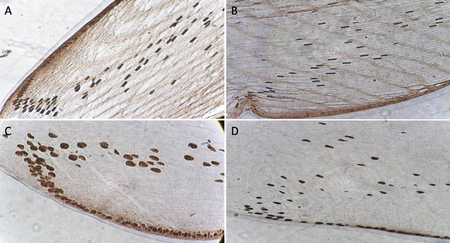
 Figure 6 of Zhang, Mol Vis 2010; 16:1754-1763.