Figure 4. Color photograph and fluorescein angiography of irises of monkeys at 23 days after laser coagulation. In monkey number 3,
moderate iris neovascularization (A) can be observed through the whole iris with obvious leakage of fluorescein (B). In monkey number 4, severe hyphema prevented the observation of iris (C and D). In monkey number 5, no obvious iris neovascularization (E) can be observed, while IFA revealed high fluorescence at the margin of pupil and point posterior synechiae (F). In monkey number 6, thin iris neovascularization can be observed through the whole iris together with an irregular and
fixed pupil. Ectropion uveae and hemorrhages adhering to the anterior surface of lens were present (G).
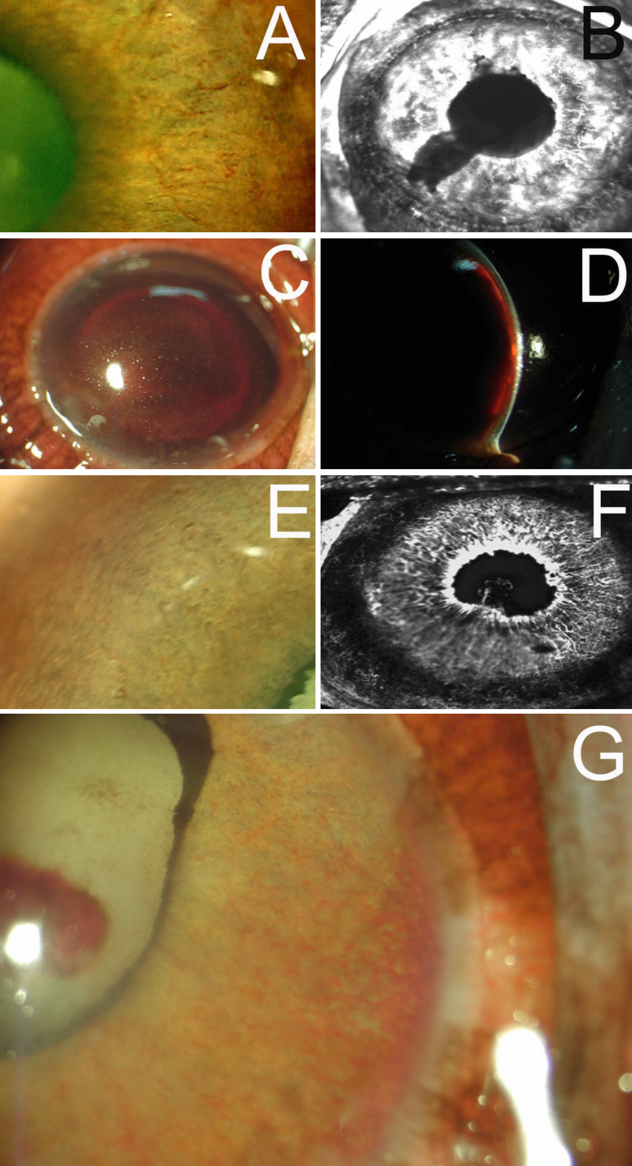
 Figure 4 of
Yuan, Mol Vis 2010; 16:1743-1753.
Figure 4 of
Yuan, Mol Vis 2010; 16:1743-1753.