Figure 4. Analysis of spheres and their
progeny. A: Immunocytochemical analysis of sphere colonies on
day 7. Bright-field images and immunostaining of spheres for p63 (an
epidermal stem/progenitor cell marker), p75NTR (an epidermal
basal progenitor cell marker), cytokeratins 3 and 12 (differentiated
epithelial cell markers, nestin (a neural stem cell marker),
microtubule-associated protein 2 (MAP2: a differentiated neuronal cell
marker), and neuron-specific enolase (NSE: a differentiated neuronal
cell marker). Each colony has been labeled by BrdU. As a control, IgG
was used instead of the primary antibody. Scale bar=100 µm. B:
Double immunocytochemical staining of a sphere colony. The spheres are
double immunostained by nestin and cytokeratin 12 or by p63 and alpha
smooth muscle actin (αSMA). Scale bar=100 µm. C: RT–PCR of
corneal epithelial tissues, spheres, and their progeny. Genes for P63,
keratin
3, keratin 12, and nestin are present in corneal epithelial
tissues, spheres, and their progeny derived from the limbal or central
regions, but not in total RNA processed without reverse-transcription. D:
Immunocytochemical
analysis of differentiated cells obtained from
spheres. Cells migrating out of the spheres express both cytokeratin 3
and cytokeratin 12 (differentiated epithelial cell markers). These
cells are also positive for MAP2, and NSE. Scale bar=100 µm.
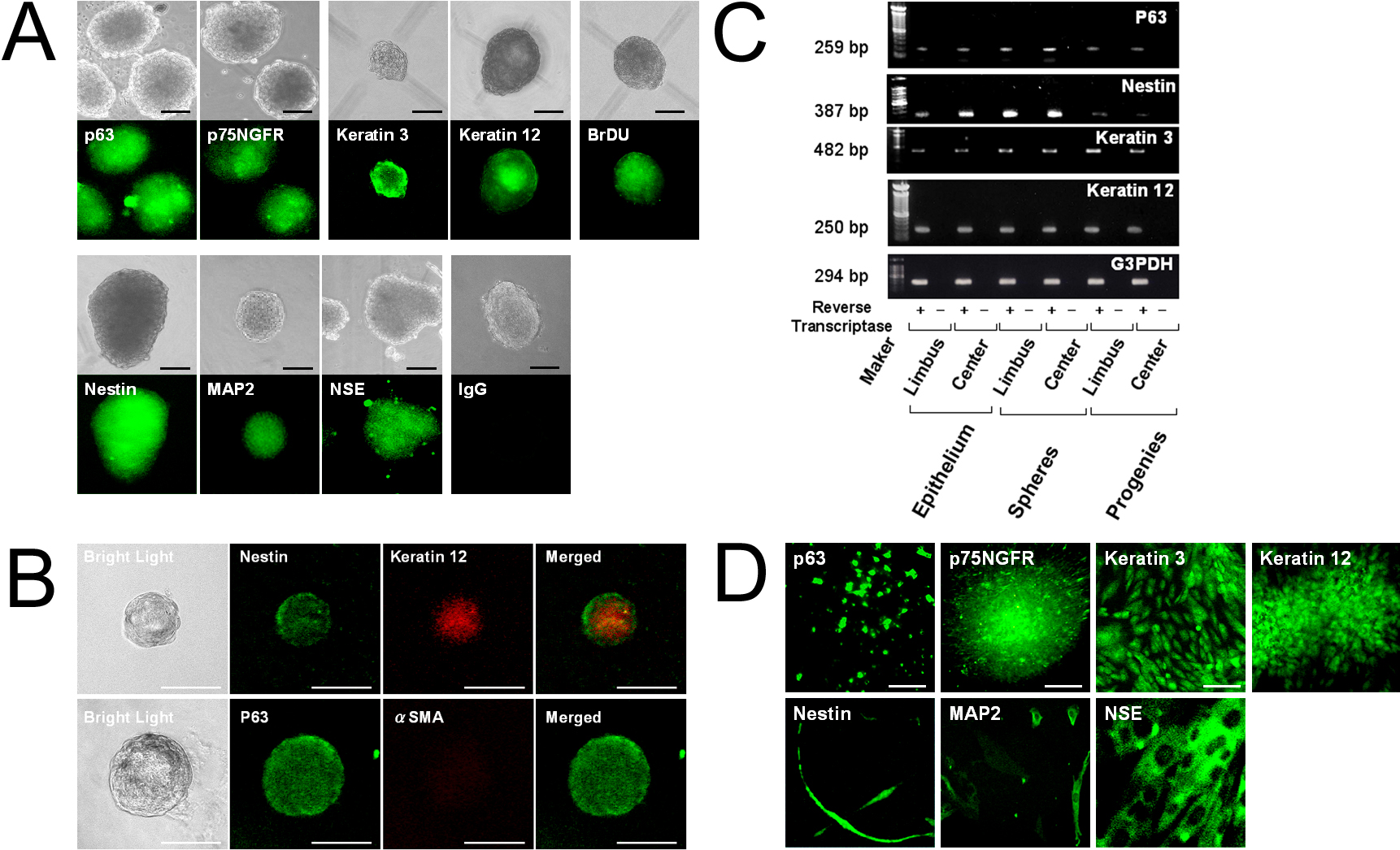
 Figure 4 of Mimura, Mol Vis 2010; 16:1712-1719.